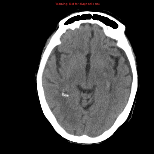
File:Bilateral subdural hemorrhage (Radiopaedia 8819-9620 Axial non-contrast 9).jpg
Jump to navigation
Jump to search
No higher resolution available.
Bilateral_subdural_hemorrhage_(Radiopaedia_8819-9620_Axial_non-contrast_9).jpg (512 × 512 pixels, file size: 81 KB, MIME type: image/jpeg)
Summary:
| Description |
|
| Date | Published: 1st Mar 2010 |
| Source | https://radiopaedia.org/cases/bilateral-subdural-haemorrhage-2 |
| Author | Jeremy Jones |
| Permission (Permission-reusing-text) |
http://creativecommons.org/licenses/by-nc-sa/3.0/ |
Licensing:
Attribution-NonCommercial-ShareAlike 3.0 Unported (CC BY-NC-SA 3.0)
File history
Click on a date/time to view the file as it appeared at that time.
| Date/Time | Thumbnail | Dimensions | User | Comment | |
|---|---|---|---|---|---|
| current | 09:29, 14 June 2021 | | 512 × 512 (81 KB) | Fæ (talk | contribs) | Radiopaedia project rID:8819 (batch #4341-9 A9) |
You cannot overwrite this file.
File usage
There are no pages that use this file.
.jpg&oldid=543782)