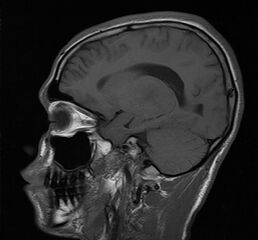
File:Bilateral thalamic gliomata (Radiopaedia 42538-45664 Sagittal T1 14).jpg
Jump to navigation
Jump to search

Size of this preview: 645 × 599 pixels. Other resolutions: 258 × 240 pixels | 517 × 480 pixels | 792 × 736 pixels.
Original file (792 × 736 pixels, file size: 70 KB, MIME type: image/jpeg)
Summary:
| Description |
|
| Date | Published: 14th Feb 2016 |
| Source | https://radiopaedia.org/cases/bilateral-thalamic-gliomata |
| Author | Ramy El Kolali |
| Permission (Permission-reusing-text) |
http://creativecommons.org/licenses/by-nc-sa/3.0/ |
Licensing:
Attribution-NonCommercial-ShareAlike 3.0 Unported (CC BY-NC-SA 3.0)
File history
Click on a date/time to view the file as it appeared at that time.
| Date/Time | Thumbnail | Dimensions | User | Comment | |
|---|---|---|---|---|---|
| current | 18:05, 14 June 2021 | | 792 × 736 (70 KB) | Fæ (talk | contribs) | Radiopaedia project rID:42538 (batch #4368-114 F14) |
You cannot overwrite this file.
File usage
There are no pages that use this file.
.jpg&oldid=547191)