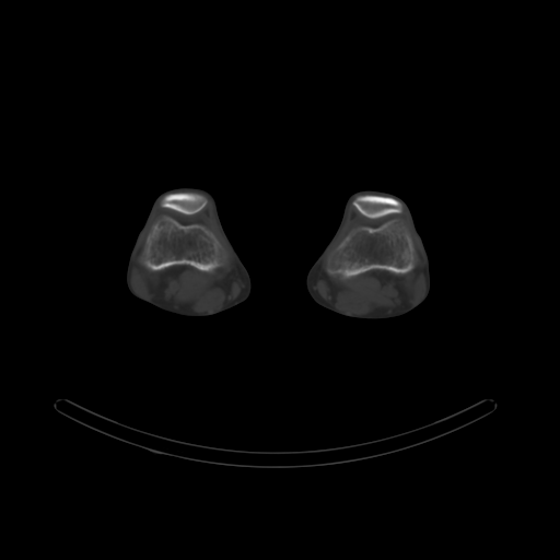
File:Bilateral tibial insufficiency fractures (Radiopaedia 46212-50612 Axial bone window 51).png
Jump to navigation
Jump to search
Bilateral_tibial_insufficiency_fractures_(Radiopaedia_46212-50612_Axial_bone_window_51).png (512 × 512 pixels, file size: 13 KB, MIME type: image/png)
Summary:
| Description |
|
| Date | Published: 3rd Jul 2016 |
| Source | https://radiopaedia.org/cases/bilateral-tibial-insufficiency-fractures |
| Author | Bruno Di Muzio |
| Permission (Permission-reusing-text) |
http://creativecommons.org/licenses/by-nc-sa/3.0/ |
Licensing:
Attribution-NonCommercial-ShareAlike 3.0 Unported (CC BY-NC-SA 3.0)
File history
Click on a date/time to view the file as it appeared at that time.
| Date/Time | Thumbnail | Dimensions | User | Comment | |
|---|---|---|---|---|---|
| current | 18:39, 14 June 2021 |  | 512 × 512 (13 KB) | Fæ (talk | contribs) | Radiopaedia project rID:46212 (batch #4375-83 B51) |
You cannot overwrite this file.
File usage
The following page uses this file:
