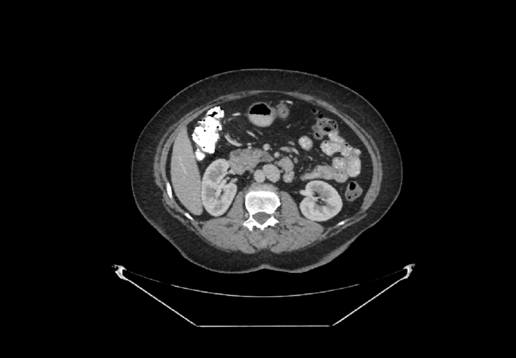
File:Bilateral urolithiasis with incidentally detected splenic artery aneurysm and left inferior vena cava (Radiopaedia 44467-48127 B 44).jpg
Jump to navigation
Jump to search
Size of this preview: 800 × 555 pixels. Other resolutions: 320 × 222 pixels | 640 × 444 pixels | 1,024 × 710 pixels.
Original file (1,024 × 710 pixels, file size: 126 KB, MIME type: image/jpeg)
Summary:
| Description |
|
| Date | Published: 23rd Apr 2016 |
| Source | https://radiopaedia.org/cases/bilateral-urolithiasis-with-incidentally-detected-splenic-artery-aneurysm-and-left-inferior-vena-cava |
| Author | Essam G Ghonaim |
| Permission (Permission-reusing-text) |
http://creativecommons.org/licenses/by-nc-sa/3.0/ |
Licensing:
Attribution-NonCommercial-ShareAlike 3.0 Unported (CC BY-NC-SA 3.0)
File history
Click on a date/time to view the file as it appeared at that time.
| Date/Time | Thumbnail | Dimensions | User | Comment | |
|---|---|---|---|---|---|
| current | 00:13, 15 June 2021 | | 1,024 × 710 (126 KB) | Fæ (talk | contribs) | Radiopaedia project rID:44467 (batch #4394-89 B44) |
You cannot overwrite this file.
File usage
There are no pages that use this file.
.jpg&oldid=549436)