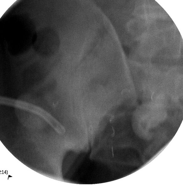
File:Bladder exstrophy and ileal conduit stones (Radiopaedia 35761-37310 scout 1).JPG
Jump to navigation
Jump to search

Size of this preview: 583 × 599 pixels. Other resolutions: 233 × 240 pixels | 467 × 480 pixels | 747 × 768 pixels | 1,097 × 1,128 pixels.
Original file (1,097 × 1,128 pixels, file size: 135 KB, MIME type: image/jpeg)
Summary:
| Description |
|
| Date | 23 Apr 2015 |
| Source | Bladder exstrophy and ileal conduit stones |
| Author | Jayanth Keshavamurthy |
| Permission (Permission-reusing-text) |
http://creativecommons.org/licenses/by-nc-sa/3.0/ |
Licensing:
Attribution-NonCommercial-ShareAlike 3.0 Unported (CC BY-NC-SA 3.0)
| This file is ineligible for copyright and therefore in the public domain, because it is a technical image created as part of a standard medical diagnostic procedure. No creative element rising above the threshold of originality was involved in its production.
|
File history
Click on a date/time to view the file as it appeared at that time.
| Date/Time | Thumbnail | Dimensions | User | Comment | |
|---|---|---|---|---|---|
| current | 04:10, 16 June 2021 |  | 1,097 × 1,128 (135 KB) | Fæ (talk | contribs) | Radiopaedia project rID:35761 (batch #4578-1 A1) |
You cannot overwrite this file.
File usage
There are no pages that use this file.
