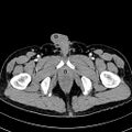
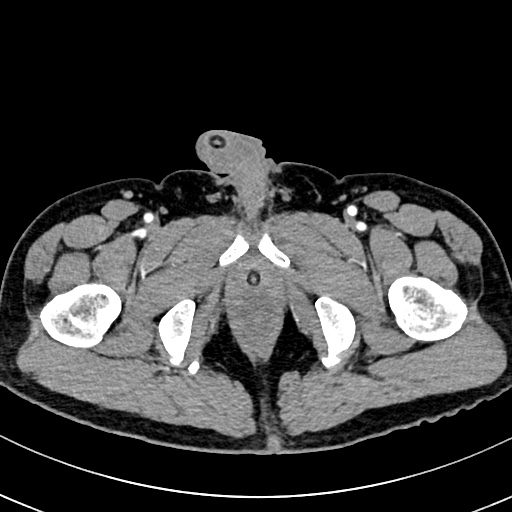
File:Bladder leiomyoma (Radiopaedia 76700-88518 Axial C+ Urogram 349).jpg

From NC Commons
Jump to navigation Jump to search

Bladder_leiomyoma_(Radiopaedia_76700-88518_Axial_C+_Urogram_349).jpg(512 × 512 pixels, file size: 72 KB, MIME type: image/jpeg)

Summary:

Description
  • Radiopaedia case ID: 76700
  • Image ID: 52481831
  • Image stack position: 349/399
  • Plane projection: Axial
  • Aux modality: C+ Urogram
  • Study description: CT Urography
  • Modality: CT
  • System: Urogenital
  • Findings: The urinary bladder is adequately distended with mild circumferential wall thickening. There is a heterogeneously enhancing, well-circumscribed soft tissue mass projecting into the right anterolateral portion of the bladder lumen measuring approximately 4.5 x 3.9 x 4.2 cm (AP x W x CC). Surrounding perivesical stranding densities are seen. Intravesical air locules (from recent instrumentation) and Foley catheter balloon are likewise identified. The prostate gland is enlarged. There is a slightly enlarged lymph node in the left pelvic sidewall /internal iliac chain. No ascites. Subcentimeter renal cysts (Bosniak I) are seen. The kidneys are normal in size and contour. No hydronephrosis or opaque lithiasis. Incidental note of cholelithiases without signs of inflammation as well as small non-inflamed diverticulum in the cecum.
Date Published: 29th Apr 2020
Source https://radiopaedia.org/cases/bladder-leiomyoma
Author Jerald Garvin Lim
Permission
(Permission-reusing-text)
http://creativecommons.org/licenses/by-nc-sa/3.0/

Licensing:

Attribution-NonCommercial-ShareAlike 3.0 Unported (CC BY-NC-SA 3.0)

File history

Click on a date/time to view the file as it appeared at that time.

Date/TimeThumbnailDimensionsUserComment
current06:17, 16 June 2021Thumbnail for version as of 06:17, 16 June 2021512 × 512 (72 KB) (talk | contribs)Radiopaedia project rID:76700 (batch #4586-680 B349)