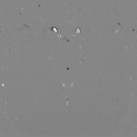
File:Blake's pouch cyst (Radiopaedia 75680-87010 Axial 26).jpg
Jump to navigation
Jump to search
Blake's_pouch_cyst_(Radiopaedia_75680-87010_Axial_26).jpg (460 × 460 pixels, file size: 83 KB, MIME type: image/jpeg)
Summary:
| Description |
|
| Date | Published: 6th Apr 2020 |
| Source | https://radiopaedia.org/cases/blakes-pouch-cyst-6 |
| Author | Dalia Ibrahim |
| Permission (Permission-reusing-text) |
http://creativecommons.org/licenses/by-nc-sa/3.0/ |
Licensing:
Attribution-NonCommercial-ShareAlike 3.0 Unported (CC BY-NC-SA 3.0)
File history
Click on a date/time to view the file as it appeared at that time.
| Date/Time | Thumbnail | Dimensions | User | Comment | |
|---|---|---|---|---|---|
| current | 15:31, 16 June 2021 |  | 460 × 460 (83 KB) | Fæ (talk | contribs) | Radiopaedia project rID:75680 (batch #4617-286 H26) |
You cannot overwrite this file.
File usage
The following page uses this file:
