File:Bleeding duodenal ulcer (Radiopaedia 53244-59213 C 29).png

From NC Commons
Jump to navigation Jump to search

Bleeding_duodenal_ulcer_(Radiopaedia_53244-59213_C_29).png(512 × 512 pixels, file size: 78 KB, MIME type: image/png)

Summary:

Description
  • Radiopaedia case ID: 53244
  • Image ID: 30269866
  • Image stack position: 29/60
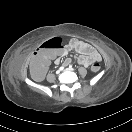
  • Plane projection: Axial
  • Aux modality: C+ portal venous phase
  • Study description: CT Abdomen and Pelvis (GI bleed protocol)
  • Modality: CT
  • System: Gastrointestinal
  • Findings: There is active contrast blush within the second portion of the duodenum associated with diffuse duodenal wall thickening. No other hemorrhagic foci identified. Bowel has otherwise unremarkable appearances, with normal mural enhancement. No free fluid or free gas in the peritoneal cavity. Focal metallic surgical material within the right colon posterior wall. Apart from a simple cyst in the segment VII, liver has normal appearances. The gallbladder, pancreas, adrenal glands, spleen, and kidneys appear unremarkable. Left femoral venous catheter in situ with the tip in the left common iliac vein. Diffuse subcutaneous edema (anasarca). No suspicious bone lesions. Bilateral pleural effusions, with loculated appearances on the right.
Date Published: 29th May 2017
Source https://radiopaedia.org/cases/bleeding-duodenal-ulcer
Author Bruno Di Muzio
Permission
(Permission-reusing-text)
http://creativecommons.org/licenses/by-nc-sa/3.0/

Licensing:

Attribution-NonCommercial-ShareAlike 3.0 Unported (CC BY-NC-SA 3.0)

File history

Click on a date/time to view the file as it appeared at that time.

Date/TimeThumbnailDimensionsUserComment
current15:55, 16 June 2021Thumbnail for version as of 15:55, 16 June 2021512 × 512 (78 KB) (talk | contribs)Radiopaedia project rID:53244 (batch #4620-109 C29)

The following page uses this file:

Metadata