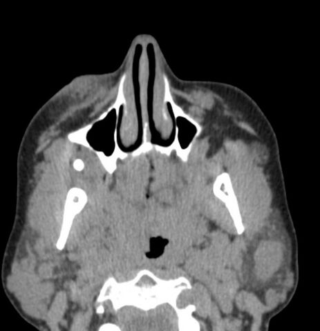
File:Blue rubber bleb nevus syndrome (Radiopaedia 51171-56823 A 27).jpg
Jump to navigation
Jump to search

Size of this preview: 578 × 600 pixels. Other resolutions: 231 × 240 pixels | 463 × 480 pixels | 671 × 696 pixels.
Original file (671 × 696 pixels, file size: 37 KB, MIME type: image/jpeg)
Summary:
| Description |
|
| Date | Published: 7th Feb 2017 |
| Source | https://radiopaedia.org/cases/blue-rubber-bleb-naevus-syndrome |
| Author | Yair Glick |
| Permission (Permission-reusing-text) |
http://creativecommons.org/licenses/by-nc-sa/3.0/ |
Licensing:
Attribution-NonCommercial-ShareAlike 3.0 Unported (CC BY-NC-SA 3.0)
File history
Click on a date/time to view the file as it appeared at that time.
| Date/Time | Thumbnail | Dimensions | User | Comment | |
|---|---|---|---|---|---|
| current | 00:40, 17 June 2021 | | 671 × 696 (37 KB) | Fæ (talk | contribs) | Radiopaedia project rID:51171 (batch #4678-27 A27) |
You cannot overwrite this file.
File usage
The following page uses this file:
.jpg&oldid=566199)