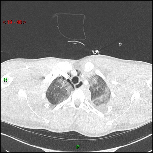
File:Blunt thoracic trauma (Radiopaedia 73454-84221 Axial lung window 17).jpg

From NC Commons
Jump to navigation Jump to search

Blunt_thoracic_trauma_(Radiopaedia_73454-84221_Axial_lung_window_17).jpg(512 × 512 pixels, file size: 75 KB, MIME type: image/jpeg)

Summary:

Description
  • Radiopaedia case ID: 73454
  • Image ID: 51918819
  • Image stack position: 17/100
  • Plane projection: Axial
  • Aux modality: lung window
  • Study findings: Partially confluent ground glass changes and centrilobular nodules predominantly involving the left lung presumed mostly secondary to contusional injury. Pneumopericardium with sleeve of dependent high density fluid in the lower left aspect of the sac (coronal reformats). Pneumomediastinum tracking up to the anterior jugular veins. Small bilateral pneumothoraces are present. Shallow left hemothorax. Prominence of the prevertebral and periaortic planes, without evidence of aortic rupture. Multilevel traumatic disruption of posterior ribs and vertebral transverse processes on the left side.
  • Modality: CT
  • System: Cardiac
  • Findings: Tiny right apical pneumothorax. Haziness over mid and lower zones of the left lung. Subtle lucency over left heart border. Some left-sided posterior rib fractures are visible. Asymmetric soft tissue density over the left clavicular region.
Date Published: 14th Jan 2020
Source https://radiopaedia.org/cases/blunt-thoracic-trauma
Author Umberto Pisano
Permission
(Permission-reusing-text)
http://creativecommons.org/licenses/by-nc-sa/3.0/

Licensing:

Attribution-NonCommercial-ShareAlike 3.0 Unported (CC BY-NC-SA 3.0)

File history

Click on a date/time to view the file as it appeared at that time.

Date/TimeThumbnailDimensionsUserComment
current07:51, 17 June 2021Thumbnail for version as of 07:51, 17 June 2021512 × 512 (75 KB) (talk | contribs)Radiopaedia project rID:73454 (batch #4686-17 A17)

Metadata