File:Boerhaave syndrome (Radiopaedia 39382-41661 A 61).png

From NC Commons
Jump to navigation Jump to search

Boerhaave_syndrome_(Radiopaedia_39382-41661_A_61).png(512 × 512 pixels, file size: 196 KB, MIME type: image/png)

Summary:

Description
  • Radiopaedia case ID: 39382
  • Image ID: 15663834
  • Image stack position: 61/65
  • Plane projection: Axial
  • Aux modality: Post oral contrast
  • Study description: CT with oral contrast
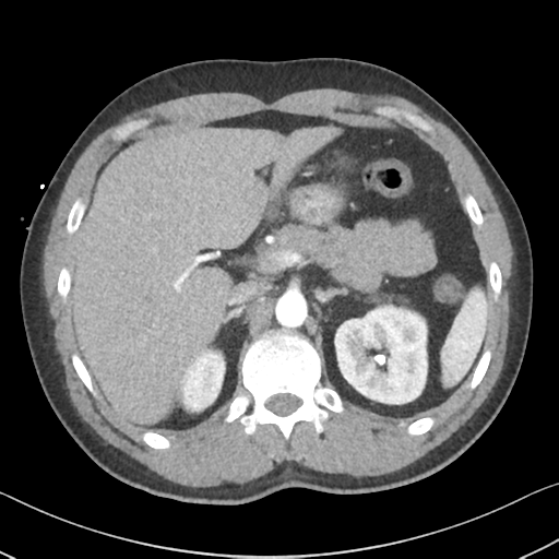
  • Study findings: Arterial phase with oral contrast (misprotocoled - should have been without IV contrast). Once again the extensive pneumomediastinum is noted. Bilateral pleural effusions and consolidation is noted. There is hold-up of oral contrast within the esophagus. There is a large amount of contrast in the posterior mediastinum surrounding and a small amount of contrast noted extending into the left pleural cavity. A discrete point of leak is suspected posteriorly just superior to the hiatus hernia. The upper abdominal contents are unremarkable. Conclusion Boerhaave syndrome with contrast extravasation into the posterior mediastinum and left hemithorax.
  • Modality: CT
  • System: Gastrointestinal
  • Findings: There is a veiling opacity over the left hemithorax, likely from pleural fluid on the supine film. There is a retrocardiac opacity that obscures the diaphragmatic silhouette, indicating left lower lobe consolidation.
Date Published: 4th Sep 2015
Source https://radiopaedia.org/cases/boerhaave-syndrome-9
Author Craig Hacking
Permission
(Permission-reusing-text)
http://creativecommons.org/licenses/by-nc-sa/3.0/

Licensing:

Attribution-NonCommercial-ShareAlike 3.0 Unported (CC BY-NC-SA 3.0)

File history

Click on a date/time to view the file as it appeared at that time.

Date/TimeThumbnailDimensionsUserComment
current13:18, 17 June 2021Thumbnail for version as of 13:18, 17 June 2021512 × 512 (196 KB) (talk | contribs)Radiopaedia project rID:39382 (batch #4704-61 A61)

The following page uses this file:

Metadata