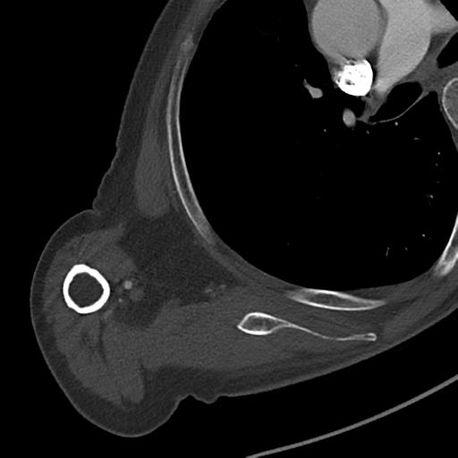
File:Bone metastasis from squamous cell carcinoma (Radiopaedia 30133-30730 A 19).jpg

From NC Commons
Jump to navigation Jump to search

Bone_metastasis_from_squamous_cell_carcinoma_(Radiopaedia_30133-30730_A_19).jpg(512 × 512 pixels, file size: 22 KB, MIME type: image/jpeg)

Summary:

Description
  • Radiopaedia case ID: 30133
  • Image ID: 7661501
  • Image stack position: 19/62
  • Plane projection: RT SHOULDER AX THICK BONE
  • Study findings: A large ( 89 x 110 x 79 mm ) soft tissue mass replaces most of the superior right scapula with a pathological fracture through the base of the acromium and a further fracture through the body of the scapula. Mass extends to the margin of the glenohumeral joint but not into it. There is no involvement of the adjacent ribs or right humeral head. The mass does not encase the adjacent axillary artery or vein. Surgical staples along the right side of the neck and an absent right sternocleidomastoid in keeping with the history of previous oropharyngeal cancer. A 59 x 66 x 95 mm hypodense lesion in segment VI of the liver has multiple small satellite nodules  and there is stranding in the surrounding fat. There is a linear hypodensity extending from the lesion to the posterior division of the right portal vein that is suspicious for thrombus in the segmental portal vein. There are multiple enlarged porta hepatis lymph nodes measuring up to 17 x 26 mm. Multiple calcified gallstones are noted. The spleen, pancreas, kidneys and adrenals are unremarkable. The prostate is enlarged ( 59 x 47 x 30 mm ). There is moderate atheroma in the abdominal aorta. Conclusion Large right scapula destructive lesion with an associated soft tissue mass. In this setting the lesion likely represents a metastatic deposit. Possible further lesion in the right ilium. Large liver lesion with surrounding satellite lesions and possible portal segmental vein thrombosis and associated lymphadenopathy.
  • Modality: CT
  • System: Oncology
  • Findings: Fracture lines are seen through the blade and spine of the right scapula.  A destructive lytic lesion of the spine of scapula is best seen on the lateral projection but the length of the spine is also absent on the AP film. Appearance is suspicious for a pathological fracture. Surgical clips are noted in the right base of neck/thoracic apex. Old right-sided rib fractures.
Date Published: 2nd Aug 2014
Source https://radiopaedia.org/cases/bone-metastasis-from-squamous-cell-carcinoma
Author Jack Ren
Permission
(Permission-reusing-text)
http://creativecommons.org/licenses/by-nc-sa/3.0/

Licensing:

Attribution-NonCommercial-ShareAlike 3.0 Unported (CC BY-NC-SA 3.0)

File history

Click on a date/time to view the file as it appeared at that time.

Date/TimeThumbnailDimensionsUserComment
current23:32, 17 June 2021Thumbnail for version as of 23:32, 17 June 2021512 × 512 (22 KB) (talk | contribs)Radiopaedia project rID:30133 (batch #4759-19 A19)