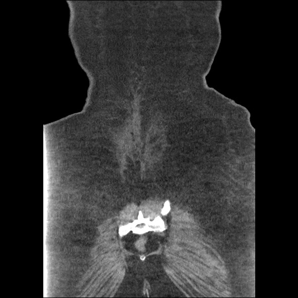
File:Bowel and splenic infarcts in acute lymphocytic leukemia (Radiopaedia 61055-68915 B 54).jpg
Jump to navigation
Jump to search
Bowel_and_splenic_infarcts_in_acute_lymphocytic_leukemia_(Radiopaedia_61055-68915_B_54).jpg (592 × 592 pixels, file size: 69 KB, MIME type: image/jpeg)
Summary:
| Description |
|
| Date | Published: 15th Jun 2018 |
| Source | https://radiopaedia.org/cases/bowel-and-splenic-infarcts-in-acute-lymphocytic-leukaemia |
| Author | Vikas Shah |
| Permission (Permission-reusing-text) |
http://creativecommons.org/licenses/by-nc-sa/3.0/ |
Licensing:
Attribution-NonCommercial-ShareAlike 3.0 Unported (CC BY-NC-SA 3.0)
File history
Click on a date/time to view the file as it appeared at that time.
| Date/Time | Thumbnail | Dimensions | User | Comment | |
|---|---|---|---|---|---|
| current | 21:21, 18 June 2021 |  | 592 × 592 (69 KB) | Fæ (talk | contribs) | Radiopaedia project rID:61055 (batch #4838-195 B54) |
You cannot overwrite this file.
File usage
The following page uses this file:
