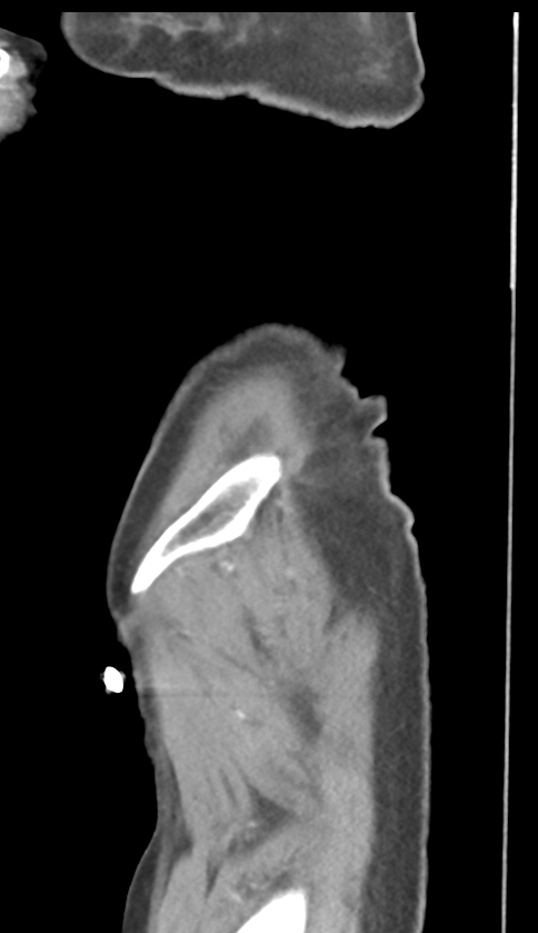
File:Bowel ischemia (Radiopaedia 58273-65382 C 8).png
Jump to navigation
Jump to search
Size of this preview: 346 × 600 pixels. Other resolutions: 138 × 240 pixels | 484 × 839 pixels.
Original file (484 × 839 pixels, file size: 76 KB, MIME type: image/png)
Summary:
| Description |
|
| Date | Published: 11th Feb 2018 |
| Source | https://radiopaedia.org/cases/bowel-ischaemia-4 |
| Author | Heather Pascoe |
| Permission (Permission-reusing-text) |
http://creativecommons.org/licenses/by-nc-sa/3.0/ |
Licensing:
Attribution-NonCommercial-ShareAlike 3.0 Unported (CC BY-NC-SA 3.0)
File history
Click on a date/time to view the file as it appeared at that time.
| Date/Time | Thumbnail | Dimensions | User | Comment | |
|---|---|---|---|---|---|
| current | 00:04, 19 June 2021 | | 484 × 839 (76 KB) | Fæ (talk | contribs) | Radiopaedia project rID:58273 (batch #4844-140 C8) |
You cannot overwrite this file.
File usage
The following page uses this file:
.png&oldid=584344)