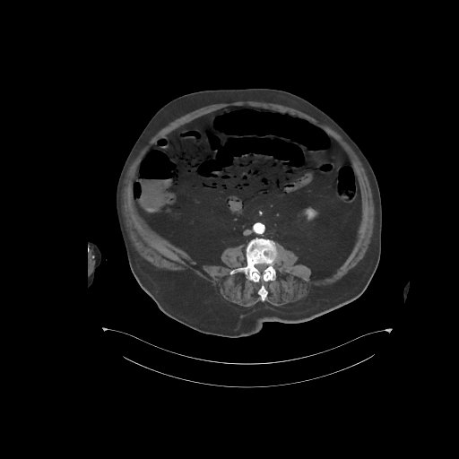
File:Bowel ischemia secondary to SMA occlusion with extensive portomesenteric venous gas (Radiopaedia 54656-60871 A 30).jpg
Jump to navigation
Jump to search
No higher resolution available.
Bowel_ischemia_secondary_to_SMA_occlusion_with_extensive_portomesenteric_venous_gas_(Radiopaedia_54656-60871_A_30).jpg (512 × 512 pixels, file size: 38 KB, MIME type: image/jpeg)
Summary:
| Description |
|
| Date | Published: 20th Jul 2017 |
| Source | https://radiopaedia.org/cases/bowel-ischaemia-secondary-to-sma-occlusion-with-extensive-portomesenteric-venous-gas |
| Author | Benedikt Beilstein |
| Permission (Permission-reusing-text) |
http://creativecommons.org/licenses/by-nc-sa/3.0/ |
Licensing:
Attribution-NonCommercial-ShareAlike 3.0 Unported (CC BY-NC-SA 3.0)
File history
Click on a date/time to view the file as it appeared at that time.
| Date/Time | Thumbnail | Dimensions | User | Comment | |
|---|---|---|---|---|---|
| current | 00:23, 19 June 2021 | | 512 × 512 (38 KB) | Fæ (talk | contribs) | Radiopaedia project rID:54656 (batch #4845-30 A30) |
You cannot overwrite this file.
File usage
The following page uses this file:
.jpg&oldid=584440)