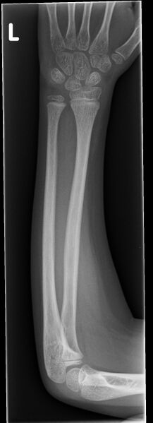
File:Bowing fracture radius and ulna (Radiopaedia 44173-47759 Frontal 1).jpg
Jump to navigation
Jump to search

Size of this preview: 217 × 599 pixels. Other resolutions: 87 × 240 pixels | 174 × 480 pixels | 873 × 2,409 pixels.
Original file (873 × 2,409 pixels, file size: 403 KB, MIME type: image/jpeg)
Summary:
| Description |
|
| Date | Published: 16th May 2016 |
| Source | https://radiopaedia.org/cases/bowing-fracture-radius-and-ulna |
| Author | Jeremy Jones |
| Permission (Permission-reusing-text) |
http://creativecommons.org/licenses/by-nc-sa/3.0/ |
Licensing:
Attribution-NonCommercial-ShareAlike 3.0 Unported (CC BY-NC-SA 3.0)
File history
Click on a date/time to view the file as it appeared at that time.
| Date/Time | Thumbnail | Dimensions | User | Comment | |
|---|---|---|---|---|---|
| current | 04:54, 19 June 2021 | 873 × 2,409 (403 KB) | Fæ (talk | contribs) | Radiopaedia project rID:44173 (batch #4856-1 A1) |
You cannot overwrite this file.
File usage
There are no pages that use this file.
.jpg&oldid=586109)