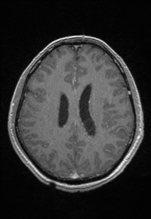
File:Brain abscess with ventriculitis (Radiopaedia 85703-101495 Axial T1 C+ 116).jpg
Jump to navigation
Jump to search
Size of this preview: 412 × 600 pixels. Other resolutions: 165 × 240 pixels | 500 × 728 pixels.
Original file (500 × 728 pixels, file size: 125 KB, MIME type: image/jpeg)
Summary:
| Description |
|
| Date | Published: 10th Jan 2021 |
| Source | https://radiopaedia.org/cases/brain-abscess-with-ventriculitis-3 |
| Author | Luu Hanh |
| Permission (Permission-reusing-text) |
http://creativecommons.org/licenses/by-nc-sa/3.0/ |
Licensing:
Attribution-NonCommercial-ShareAlike 3.0 Unported (CC BY-NC-SA 3.0)
File history
Click on a date/time to view the file as it appeared at that time.
| Date/Time | Thumbnail | Dimensions | User | Comment | |
|---|---|---|---|---|---|
| current | 00:54, 20 June 2021 | | 500 × 728 (125 KB) | Fæ (talk | contribs) | Radiopaedia project rID:85703 (batch #4947-140 B116) |
You cannot overwrite this file.
File usage
The following page uses this file:
.jpg&oldid=591762)