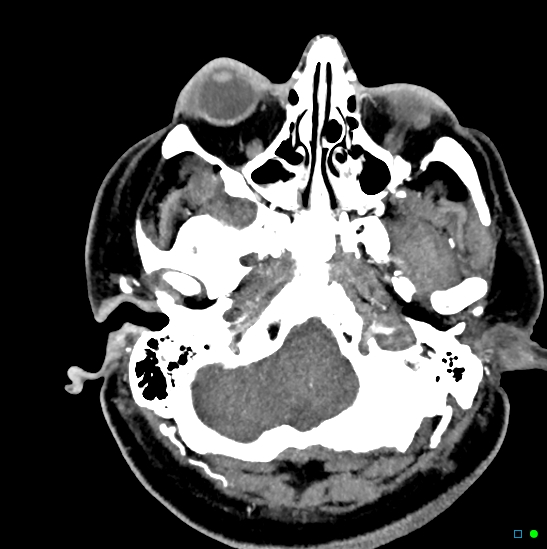
File:Brain death on MRI and CT angiography (Radiopaedia 42560-45841 B 44).jpg
Jump to navigation
Jump to search
No higher resolution available.
Brain_death_on_MRI_and_CT_angiography_(Radiopaedia_42560-45841_B_44).jpg (547 × 549 pixels, file size: 127 KB, MIME type: image/jpeg)
Summary:
| Description |
|
| Date | Published: 28th Feb 2016 |
| Source | https://radiopaedia.org/cases/brain-death-on-mri-and-ct-angiography |
| Author | Chris O'Donnell |
| Permission (Permission-reusing-text) |
http://creativecommons.org/licenses/by-nc-sa/3.0/ |
Licensing:
Attribution-NonCommercial-ShareAlike 3.0 Unported (CC BY-NC-SA 3.0)
File history
Click on a date/time to view the file as it appeared at that time.
| Date/Time | Thumbnail | Dimensions | User | Comment | |
|---|---|---|---|---|---|
| current | 07:59, 20 June 2021 | | 547 × 549 (127 KB) | Fæ (talk | contribs) | Radiopaedia project rID:42560 (batch #4963-45 B44) |
You cannot overwrite this file.
File usage
The following page uses this file:
.jpg&oldid=594265)