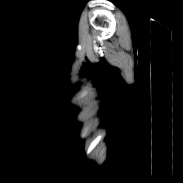
File:Brain metastases from lung cancer (Radiopaedia 27849-28093 F 4).jpg
Jump to navigation
Jump to search

Size of this preview: 600 × 600 pixels. Other resolutions: 240 × 240 pixels | 611 × 611 pixels.
Original file (611 × 611 pixels, file size: 34 KB, MIME type: image/jpeg)
Summary:
| Description |
|
| Date | Published: 22nd Feb 2014 |
| Source | https://radiopaedia.org/cases/brain-metastases-from-lung-cancer-1 |
| Author | David Cuete |
| Permission (Permission-reusing-text) |
http://creativecommons.org/licenses/by-nc-sa/3.0/ |
Licensing:
Attribution-NonCommercial-ShareAlike 3.0 Unported (CC BY-NC-SA 3.0)
File history
Click on a date/time to view the file as it appeared at that time.
| Date/Time | Thumbnail | Dimensions | User | Comment | |
|---|---|---|---|---|---|
| current | 14:18, 20 June 2021 | | 611 × 611 (34 KB) | Fæ (talk | contribs) | Radiopaedia project rID:27849 (batch #4976-248 F4) |
You cannot overwrite this file.
File usage
The following page uses this file:
.jpg&oldid=596719)