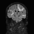
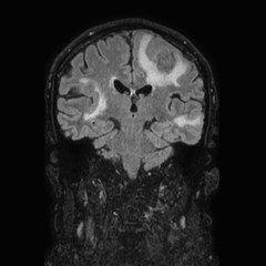
File:Brain metastases from lung cancer (Radiopaedia 83839-99028 Coronal FLAIR 46).jpg
Jump to navigation
Jump to search
Brain_metastases_from_lung_cancer_(Radiopaedia_83839-99028_Coronal_FLAIR_46).jpg (240 × 240 pixels, file size: 11 KB, MIME type: image/jpeg)
Summary:
| Description |
|
| Date | Published: 10th Nov 2020 |
| Source | https://radiopaedia.org/cases/brain-metastases-from-lung-cancer-2 |
| Author | Mohamed Abdelrazik Elsayed |
| Permission (Permission-reusing-text) |
http://creativecommons.org/licenses/by-nc-sa/3.0/ |
Licensing:
Attribution-NonCommercial-ShareAlike 3.0 Unported (CC BY-NC-SA 3.0)
File history
Click on a date/time to view the file as it appeared at that time.
| Date/Time | Thumbnail | Dimensions | User | Comment | |
|---|---|---|---|---|---|
| current | 15:31, 20 June 2021 |  | 240 × 240 (11 KB) | Fæ (talk | contribs) | Radiopaedia project rID:83839 (batch #4978-80 B46) |
You cannot overwrite this file.
File usage
The following page uses this file:
