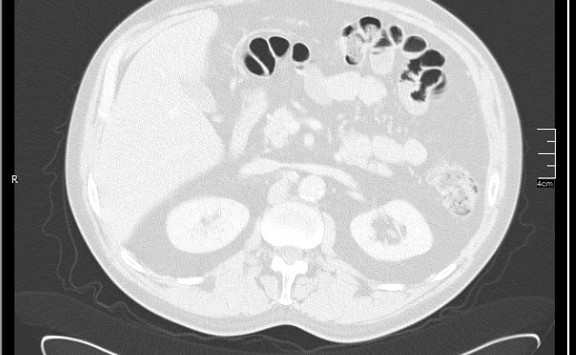
File:Brain metastases from squamocellular lung cancer (Radiopaedia 56515-63219 Axial lung window 69).jpg
Jump to navigation
Jump to search
Brain_metastases_from_squamocellular_lung_cancer_(Radiopaedia_56515-63219_Axial_lung_window_69).jpg (518 × 320 pixels, file size: 41 KB, MIME type: image/jpeg)
Summary:
| Description |
|
| Date | Published: 8th Nov 2017 |
| Source | https://radiopaedia.org/cases/brain-metastases-from-squamocellular-lung-cancer |
| Author | Dr Nikola Todorovic |
| Permission (Permission-reusing-text) |
http://creativecommons.org/licenses/by-nc-sa/3.0/ |
Licensing:
Attribution-NonCommercial-ShareAlike 3.0 Unported (CC BY-NC-SA 3.0)
File history
Click on a date/time to view the file as it appeared at that time.
| Date/Time | Thumbnail | Dimensions | User | Comment | |
|---|---|---|---|---|---|
| current | 18:25, 20 June 2021 |  | 518 × 320 (41 KB) | Fæ (talk | contribs) | Radiopaedia project rID:56515 (batch #4980-69 A69) |
You cannot overwrite this file.
File usage
The following page uses this file:
