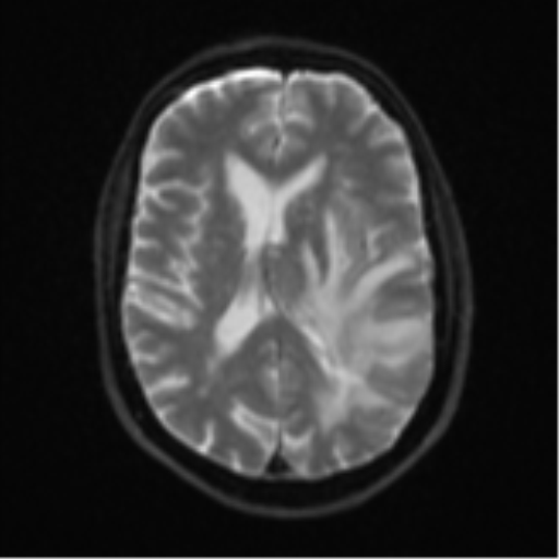
File:Brain metastasis (large cystic mass) (Radiopaedia 47497-52107 Axial DWI 16).png

From NC Commons
Jump to navigation Jump to search

Brain_metastasis_(large_cystic_mass)_(Radiopaedia_47497-52107_Axial_DWI_16).png(512 × 512 pixels, file size: 64 KB, MIME type: image/png)

Summary:

Description
  • Radiopaedia case ID: 47497
  • Image ID: 24663940
  • Image stack position: 16/56
  • Plane projection: Axial
  • Aux modality: DWI
  • Study description: MRI Brain
  • Study findings: Complex mass in the posterior aspect of the left temporal lobe is predominantly cystic with an intensely enhancing eccentric solid nodular component, which shows restricted diffusion and an elevated cerebral blood volume (CBV). No calcification was seen on the previous CT brain. The fluid component of the tumor is homogeneous and partially suppressed on FLAIR, with a thin enhancing capsule. Spectroscopy traces at the margins of the lesion do not show a choline peak or reduced NAA. Vasogenic edema surrounds the lesion and the mass effect also compresses the left cerebral peduncle and left lateral ventricle, resulting in a 6.6 mm rightward midline shift. Scattered foci of high T2/FLAIR signal abnormalities through the infra- and supratentorial white matter are nonspecific, most likely representing chronic small vessel ischemia, larger in number than expected for the patient's age group. Old pontine lacunar infarct left of midline.
  • Modality: MRI
  • System: Central Nervous System
  • Findings: Intra-axial mass lesion present within the posterior aspect of the left temporal lobe, predominantly hypoattenuating with peripheral rim enhancement, and a nodular region of enhancement along the lateral border measuring 9 mm. Diffuse white matter edema surrounding the mass, with finger-like extensions, involving the left frontal, parietal and temporal lobes. There is a resultant mass effect with partial effacement of the left lateral ventricle and a left-to-right shift of midline structures measuring 6 mm.
Date Published: 10th Sep 2016
Source https://radiopaedia.org/cases/brain-metastasis-large-cystic-mass-1
Author Bruno Di Muzio
Permission
(Permission-reusing-text)
http://creativecommons.org/licenses/by-nc-sa/3.0/

Licensing:

Attribution-NonCommercial-ShareAlike 3.0 Unported (CC BY-NC-SA 3.0)

File history

Click on a date/time to view the file as it appeared at that time.

Date/TimeThumbnailDimensionsUserComment
current03:08, 21 June 2021Thumbnail for version as of 03:08, 21 June 2021512 × 512 (64 KB) (talk | contribs)Radiopaedia project rID:47497 (batch #4992-129 E16)

Metadata