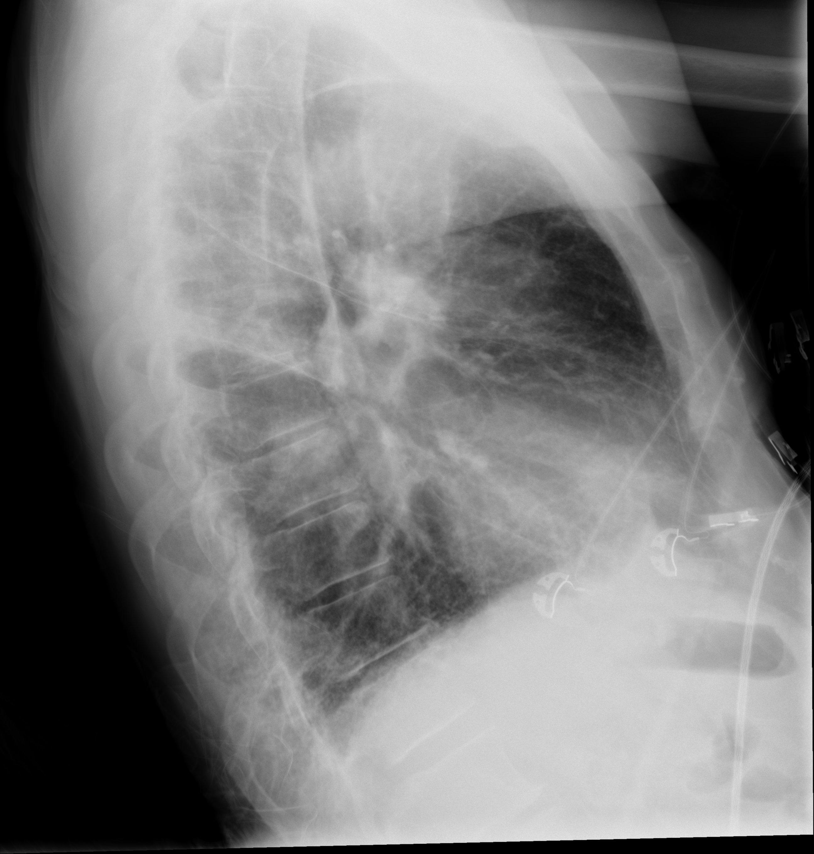
File:Brain metastasis (lung cancer) (Radiopaedia 48289-53178 Frontal-Lateral 2).png
Jump to navigation
Jump to search
Size of this preview: 571 × 599 pixels. Other resolutions: 229 × 240 pixels | 458 × 480 pixels | 732 × 768 pixels | 976 × 1,024 pixels | 1,952 × 2,048 pixels | 2,592 × 2,719 pixels.
Original file (2,592 × 2,719 pixels, file size: 2.08 MB, MIME type: image/png)
Summary:
| Description |
|
| Date | Published: 2nd Oct 2016 |
| Source | https://radiopaedia.org/cases/brain-metastasis-lung-cancer-1 |
| Author | Bruno Di Muzio |
| Permission (Permission-reusing-text) |
http://creativecommons.org/licenses/by-nc-sa/3.0/ |
Licensing:
Attribution-NonCommercial-ShareAlike 3.0 Unported (CC BY-NC-SA 3.0)
File history
Click on a date/time to view the file as it appeared at that time.
| Date/Time | Thumbnail | Dimensions | User | Comment | |
|---|---|---|---|---|---|
| current | 04:48, 21 June 2021 | | 2,592 × 2,719 (2.08 MB) | Fæ (talk | contribs) | Radiopaedia project rID:48289 (batch #4993-2 A2) |
You cannot overwrite this file.
File usage
The following page uses this file:
_(Radiopaedia_48289-53178_Frontal-Lateral_2).png&oldid=1684162)