File:Brainstem glioma (Radiopaedia 9444-10124 Sagittal T1 8).jpg

From NC Commons
Jump to navigation Jump to search

Brainstem_glioma_(Radiopaedia_9444-10124_Sagittal_T1_8).jpg(256 × 256 pixels, file size: 41 KB, MIME type: image/jpeg)

Summary:

Description
  • Radiopaedia case ID: 9444
  • Image ID: 427664
  • Image stack position: 8/11
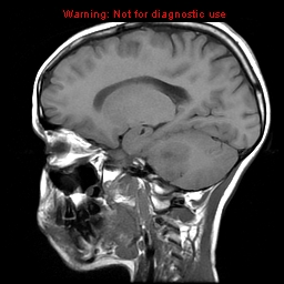
  • Plane projection: Sagittal
  • Aux modality: T1
  • Study description: MRI brain
  • Study findings: MRI of the brain confirms the presence of a large mass expanding the pons. It is T1W hypointense and T2W hyperintense, with areas on the left which demonstrate fluid signal intensity and mostly attenuated on FLAIR.  These areas also demonstrate peripheral irregular contrast enhancement. The mass overall does not demonstrate restricted diffusion, nor are there convincing areas of calcification or hemorrhage on gradient echo. The fourth ventricle is effaced and hydrocephalus is present.
  • Modality: MRI
  • System: Central Nervous System
  • Findings: Non-contrast CT of the brain demonstrates a large heterogenous mass expanding the pons, distorting the fourth ventricle and resulting in a degree of hydrocephalus. The mass is predominantly hypoattenuating centrally, with a region of relative hyperattenuation peripherally on the left.
Date Published: 19th Apr 2010
Source https://radiopaedia.org/cases/brainstem-glioma
Author Hani Makky Al Salam
Permission
(Permission-reusing-text)
http://creativecommons.org/licenses/by-nc-sa/3.0/

Licensing:

Attribution-NonCommercial-ShareAlike 3.0 Unported (CC BY-NC-SA 3.0)

File history

Click on a date/time to view the file as it appeared at that time.

Date/TimeThumbnailDimensionsUserComment
current11:18, 21 June 2021Thumbnail for version as of 11:18, 21 June 2021256 × 256 (41 KB) (talk | contribs)Radiopaedia project rID:9444 (batch #5012-8 A8)

There are no pages that use this file.