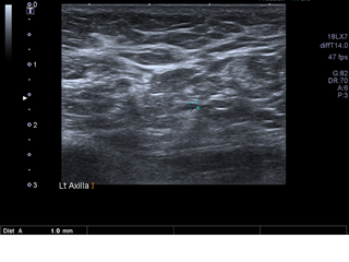
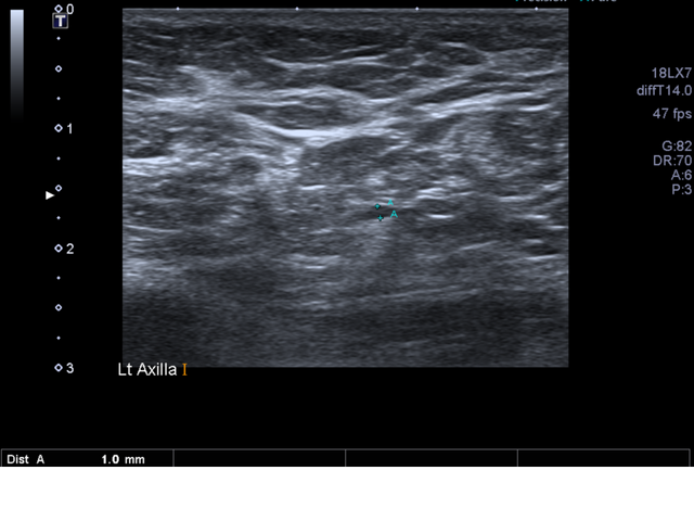
File:Breast carcinoma (Radiopaedia 45019-48981 Transverse 6).png
Jump to navigation
Jump to search

Size of this preview: 800 × 600 pixels. Other resolutions: 320 × 240 pixels | 640 × 480 pixels | 960 × 720 pixels.
Original file (960 × 720 pixels, file size: 481 KB, MIME type: image/png)
Summary:
| Description |
|
| Date | Published: 13th May 2016 |
| Source | https://radiopaedia.org/cases/breast-carcinoma-6 |
| Author | Melbourne Uni Radiology Masters |
| Permission (Permission-reusing-text) |
http://creativecommons.org/licenses/by-nc-sa/3.0/ |
Licensing:
Attribution-NonCommercial-ShareAlike 3.0 Unported (CC BY-NC-SA 3.0)
File history
Click on a date/time to view the file as it appeared at that time.
| Date/Time | Thumbnail | Dimensions | User | Comment | |
|---|---|---|---|---|---|
| current | 04:35, 22 June 2021 | | 960 × 720 (481 KB) | Fæ (talk | contribs) | Radiopaedia project rID:45019 (batch #5110-6 A6) |
You cannot overwrite this file.
File usage
There are no pages that use this file.
.png&oldid=611745)