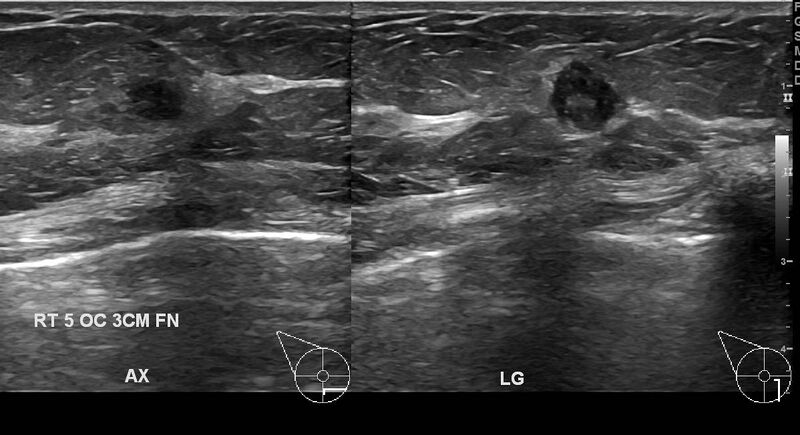
File:Breast carcinoma (multicentric multifocal in mammary Paget disease) (Radiopaedia 50966-56511 A 1).JPG
Jump to navigation
Jump to search

Size of this preview: 800 × 435 pixels. Other resolutions: 320 × 174 pixels | 640 × 348 pixels | 1,198 × 652 pixels.
Original file (1,198 × 652 pixels, file size: 101 KB, MIME type: image/jpeg)
Summary:
| Description |
|
| Date | Published: 5th Feb 2017 |
| Source | https://radiopaedia.org/cases/breast-carcinoma-multicentric-multifocal-in-mammary-paget-disease |
| Author | Nur Ahida Md Ahir |
| Permission (Permission-reusing-text) |
http://creativecommons.org/licenses/by-nc-sa/3.0/ |
Licensing:
Attribution-NonCommercial-ShareAlike 3.0 Unported (CC BY-NC-SA 3.0)
File history
Click on a date/time to view the file as it appeared at that time.
| Date/Time | Thumbnail | Dimensions | User | Comment | |
|---|---|---|---|---|---|
| current | 04:46, 22 June 2021 | | 1,198 × 652 (101 KB) | Fæ (talk | contribs) | Radiopaedia project rID:50966 (batch #5114-1 A1) |
You cannot overwrite this file.
File usage
There are no pages that use this file.
_(Radiopaedia_50966-56511_A_1).JPG&oldid=611787)