File:Breast metastases from renal cell cancer (Radiopaedia 79220-92225 C 68).jpg

From NC Commons
Jump to navigation Jump to search

Breast_metastases_from_renal_cell_cancer_(Radiopaedia_79220-92225_C_68).jpg(548 × 548 pixels, file size: 117 KB, MIME type: image/jpeg)

Summary:

Description
  • Radiopaedia case ID: 79220
  • Image ID: 52966829
  • Image stack position: 68/137
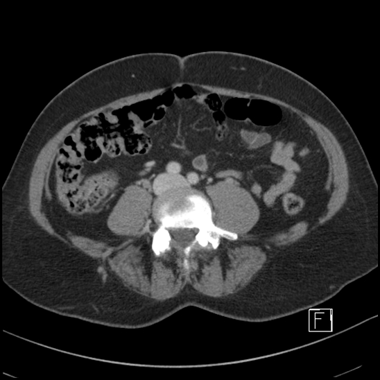
  • Plane projection: Axial
  • Aux modality: C+ portal venous phase
  • Modality: CT
  • System: Oncology
  • Findings: There are several pulmonary nodules bilaterally, the largest is in the left lower lobe measuring 17 mm. Bilateral pleural posterior basal atelectasis. No pleural thickening or pleural effusion. Heart size is normal. No pulmonary embolus identified in a non-dedicated contrast study. No lymphadenopathy within the chest. The left breast contains two peripherally dense irregular masses measuring 11 mm superiorly and 10 mm laterally. There is no axillary, supraclavicular or internal mammary lymphadenopathy. There is a large irregular peripherally enhancing centrally necrotic right renal mass which measures 90 x 79 x 99 mm (AP x LR x CC) arising from the lower pole of the kidney. Laterally there is stranding of the perirenal fat and large recruited vessels. Medially the lesion abuts the psoas major muscle without evidence of muscular invasion. No hydronephrosis. The right renal vein and IVC are clear. The left kidney contains a simple cortical cyst (42 mm). No retroperitoneal lymphadenopathy.No hepatic, parenchymal, splenic or adrenal lesion. The large and small bowel is unremarkable. The appendix incidentally contains an appendicolith. The uterus appears irregular with multiple fibroids internally. Cystic ovaries are noted bilaterally. No free fluid or free gas. Thoracic and lumbosacral alignment is normal. No vertebral metastatic lesion is evident. No pelvic or proximal femoral lesion identified. IMPRESSION Large right lower pole renal lesion with central necrosis most in keeping with a primary renal malignancy such as RCC. There is no evidence of vascular invasion or lymphadenopathy. Multiple pulmonary metastases. No osseous metastasis identified within the axial skeleton. Two peripherally dense left breast lesions may represent metastases also given the absence of lymphadenopathy. Breast US and mammography is suggested.
Date Published: 22nd Jun 2020
Source https://radiopaedia.org/cases/breast-metastases-from-renal-cell-cancer
Author Craig Hacking
Permission
(Permission-reusing-text)
http://creativecommons.org/licenses/by-nc-sa/3.0/

Licensing:

Attribution-NonCommercial-ShareAlike 3.0 Unported (CC BY-NC-SA 3.0)

File history

Click on a date/time to view the file as it appeared at that time.

Date/TimeThumbnailDimensionsUserComment
current01:06, 24 June 2021Thumbnail for version as of 01:06, 24 June 2021548 × 548 (117 KB) (talk | contribs)Radiopaedia project rID:79220 (batch #5169-250 C68)