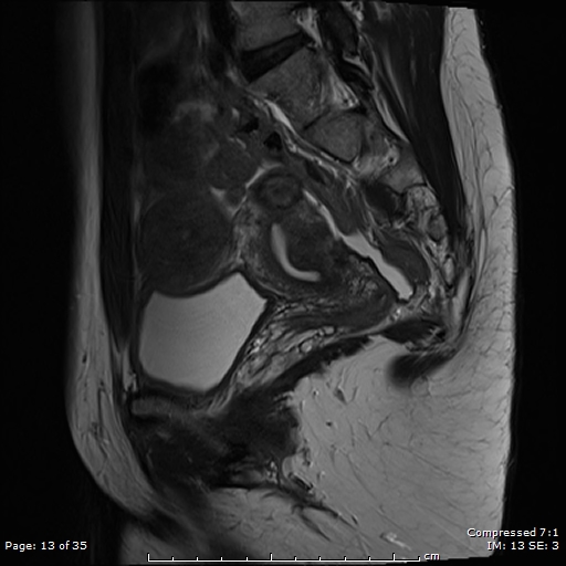
File:Bridging vessel sign - pedunculated subserosal leiomyoma (Radiopaedia 86109-102055 Sagittal T2 13).jpg
Jump to navigation
Jump to search
Bridging_vessel_sign_-_pedunculated_subserosal_leiomyoma_(Radiopaedia_86109-102055_Sagittal_T2_13).jpg (512 × 512 pixels, file size: 110 KB, MIME type: image/jpeg)
Summary:
| Description |
|
| Date | Published: 24th Jan 2021 |
| Source | https://radiopaedia.org/cases/bridging-vessel-sign-pedunculated-subserosal-leiomyoma |
| Author | Eid Kakish |
| Permission (Permission-reusing-text) |
http://creativecommons.org/licenses/by-nc-sa/3.0/ |
Licensing:
Attribution-NonCommercial-ShareAlike 3.0 Unported (CC BY-NC-SA 3.0)
File history
Click on a date/time to view the file as it appeared at that time.
| Date/Time | Thumbnail | Dimensions | User | Comment | |
|---|---|---|---|---|---|
| current | 02:52, 24 June 2021 |  | 512 × 512 (110 KB) | Fæ (talk | contribs) | Radiopaedia project rID:86109 (batch #5191-166 D13) |
You cannot overwrite this file.
File usage
There are no pages that use this file.
