File:Bronchial atresia (Radiopaedia 58271-65417 Coronal lung window 5).jpg

From NC Commons
Jump to navigation Jump to search

Bronchial_atresia_(Radiopaedia_58271-65417_Coronal_lung_window_5).jpg(512 × 412 pixels, file size: 202 KB, MIME type: image/jpeg)

Summary:

Description
  • Radiopaedia case ID: 58271
  • Image ID: 35878449
  • Image stack position: 5/53
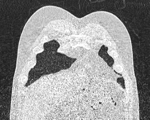
  • Plane projection: Coronal
  • Aux modality: lung window
  • Study findings: Axial lung window of pulmonary CT angiogram, shows hyperaerated and hypovascular basal segments of the right lower lobe. A mucus filled segment of dilated bronchus (bronchocele) is centrally located in the hyeprlucent lung. Note ground-glass opacities in the left lower lobe that are secondary to alveolar hemorrhage. Coronal reconstruction CT scan shows hyperinflation in the basal segments of the right lower lobe. A branching opacity is seen in the central portion of the involved segments, that represents dilated mucus-filled bronchi (bronchocele). Note ground-glass opacities in the left lower lobe that are secondary to alveolar hemorrhage that is an unrelated symptom and the cause of performing this imaging study.
  • Modality: CT
  • System: Chest
  • Findings: Supine CXR reveals a hyperlucency in the right lower zone. A central oval opacity is seen within the lucent area.
Date Published: 11th Feb 2018
Source https://radiopaedia.org/cases/bronchial-atresia-9
Author Bita Abbasi
Permission
(Permission-reusing-text)
http://creativecommons.org/licenses/by-nc-sa/3.0/

Licensing:

Attribution-NonCommercial-ShareAlike 3.0 Unported (CC BY-NC-SA 3.0)

File history

Click on a date/time to view the file as it appeared at that time.

Date/TimeThumbnailDimensionsUserComment
current13:19, 24 June 2021Thumbnail for version as of 13:19, 24 June 2021512 × 412 (202 KB) (talk | contribs)Radiopaedia project rID:58271 (batch #5235-52 B5)