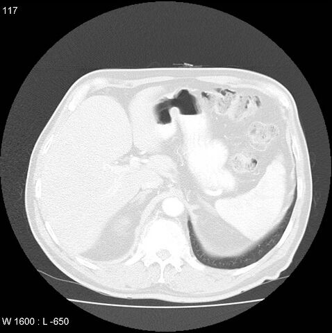
File:Bronchial carcinoid tumor with right lower lobe collapse (Radiopaedia 29060-29422 B 55).jpg
Jump to navigation
Jump to search

Size of this preview: 596 × 599 pixels. Other resolutions: 239 × 240 pixels | 478 × 480 pixels | 766 × 770 pixels.
Original file (766 × 770 pixels, file size: 118 KB, MIME type: image/jpeg)
Summary:
| Description |
|
| Date | Published: 1st May 2014 |
| Source | https://radiopaedia.org/cases/bronchial-carcinoid-tumour-with-right-lower-lobe-collapse |
| Author | Jack Ren |
| Permission (Permission-reusing-text) |
http://creativecommons.org/licenses/by-nc-sa/3.0/ |
Licensing:
Attribution-NonCommercial-ShareAlike 3.0 Unported (CC BY-NC-SA 3.0)
File history
Click on a date/time to view the file as it appeared at that time.
| Date/Time | Thumbnail | Dimensions | User | Comment | |
|---|---|---|---|---|---|
| current | 19:08, 24 June 2021 | | 766 × 770 (118 KB) | Fæ (talk | contribs) | Radiopaedia project rID:29060 (batch #5245-116 B55) |
You cannot overwrite this file.
File usage
The following page uses this file:
.jpg&oldid=620601)