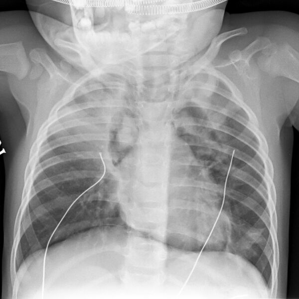
File:Bronchiolitis with pneumomediastinum (Radiopaedia 35348-36854 Frontal 1).jpg
Jump to navigation
Jump to search

Size of this preview: 600 × 600 pixels. Other resolutions: 240 × 240 pixels | 480 × 480 pixels | 768 × 768 pixels | 1,024 × 1,024 pixels | 1,800 × 1,800 pixels.
Original file (1,800 × 1,800 pixels, file size: 1.37 MB, MIME type: image/jpeg)
Summary:
| Description |
|
| Date | Published: 2nd Apr 2015 |
| Source | https://radiopaedia.org/cases/bronchiolitis-with-pneumomediastinum |
| Author | Jeremy Jones |
| Permission (Permission-reusing-text) |
http://creativecommons.org/licenses/by-nc-sa/3.0/ |
Licensing:
Attribution-NonCommercial-ShareAlike 3.0 Unported (CC BY-NC-SA 3.0)
File history
Click on a date/time to view the file as it appeared at that time.
| Date/Time | Thumbnail | Dimensions | User | Comment | |
|---|---|---|---|---|---|
| current | 03:24, 25 June 2021 |  | 1,800 × 1,800 (1.37 MB) | Fæ (talk | contribs) | Radiopaedia project rID:35348 (batch #5277-1 A1) |
You cannot overwrite this file.
File usage
There are no pages that use this file.