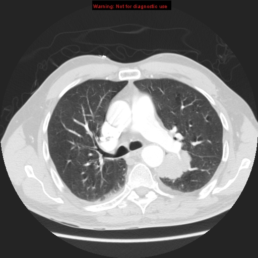
File:Bronchogenic carcinoma brain metastasis (Radiopaedia 9286-105832 Axial lung window 8).jpg
Jump to navigation
Jump to search
Bronchogenic_carcinoma_brain_metastasis_(Radiopaedia_9286-105832_Axial_lung_window_8).jpg (512 × 512 pixels, file size: 113 KB, MIME type: image/jpeg)
Summary:
| Description |
|
| Date | Published: 29th Mar 2010 |
| Source | https://radiopaedia.org/cases/bronchogenic-carcinoma-brain-metastasis |
| Author | Hani Makky Al Salam |
| Permission (Permission-reusing-text) |
http://creativecommons.org/licenses/by-nc-sa/3.0/ |
Licensing:
Attribution-NonCommercial-ShareAlike 3.0 Unported (CC BY-NC-SA 3.0)
File history
Click on a date/time to view the file as it appeared at that time.
| Date/Time | Thumbnail | Dimensions | User | Comment | |
|---|---|---|---|---|---|
| current | 08:43, 25 June 2021 |  | 512 × 512 (113 KB) | Fæ (talk | contribs) | Radiopaedia project rID:9286 (batch #5295-10 C8) |
You cannot overwrite this file.
File usage
There are no pages that use this file.
