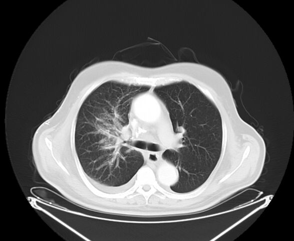
File:Bronchogenic carcinoma with lymphangitic spread (Radiopaedia 29002-29357 Axial lung window 14).jpg
Jump to navigation
Jump to search

Size of this preview: 733 × 600 pixels. Other resolutions: 293 × 240 pixels | 587 × 480 pixels | 825 × 675 pixels.
Original file (825 × 675 pixels, file size: 205 KB, MIME type: image/jpeg)
Summary:
| Description |
|
| Date | Published: 26th Apr 2014 |
| Source | https://radiopaedia.org/cases/bronchogenic-carcinoma-with-lymphangitic-spread |
| Author | Ahmed Abdrabou |
| Permission (Permission-reusing-text) |
http://creativecommons.org/licenses/by-nc-sa/3.0/ |
Licensing:
Attribution-NonCommercial-ShareAlike 3.0 Unported (CC BY-NC-SA 3.0)
File history
Click on a date/time to view the file as it appeared at that time.
| Date/Time | Thumbnail | Dimensions | User | Comment | |
|---|---|---|---|---|---|
| current | 09:10, 25 June 2021 | | 825 × 675 (205 KB) | Fæ (talk | contribs) | Radiopaedia project rID:29002 (batch #5299-15 B14) |
You cannot overwrite this file.
File usage
There are no pages that use this file.
.jpg&oldid=625965)