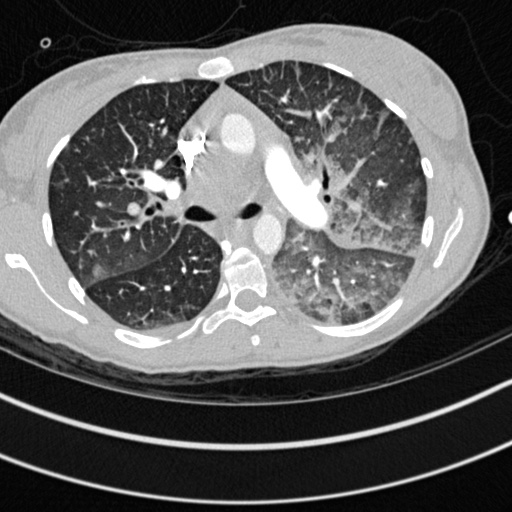
File:Bronchogenic cyst (Radiopaedia 77801-90071 Axial lung window 22).jpg
Jump to navigation
Jump to search
Bronchogenic_cyst_(Radiopaedia_77801-90071_Axial_lung_window_22).jpg (512 × 512 pixels, file size: 91 KB, MIME type: image/jpeg)
Summary:
| Description |
|
| Date | Published: 21st May 2020 |
| Source | https://radiopaedia.org/cases/bronchogenic-cyst-22 |
| Author | Annette Johnstone |
| Permission (Permission-reusing-text) |
http://creativecommons.org/licenses/by-nc-sa/3.0/ |
Licensing:
Attribution-NonCommercial-ShareAlike 3.0 Unported (CC BY-NC-SA 3.0)
File history
Click on a date/time to view the file as it appeared at that time.
| Date/Time | Thumbnail | Dimensions | User | Comment | |
|---|---|---|---|---|---|
| current | 17:44, 25 June 2021 |  | 512 × 512 (91 KB) | Fæ (talk | contribs) | Radiopaedia project rID:77801 (batch #5316-114 B22) |
You cannot overwrite this file.
File usage
The following page uses this file:
