File:Bronchogenic cyst (Radiopaedia 87717-104152 D 56).jpg

From NC Commons
Jump to navigation Jump to search

Bronchogenic_cyst_(Radiopaedia_87717-104152_D_56).jpg(512 × 481 pixels, file size: 104 KB, MIME type: image/jpeg)

Summary:

Description
  • Radiopaedia case ID: 87717
  • Image ID: 54607936
  • Image stack position: 56/96
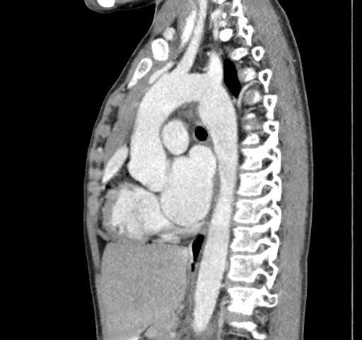
  • Plane projection: Sagittal
  • Aux modality: C+ arterial phase
  • Modality: CT
  • System: Chest
  • Findings: Large pure thin-walled fluid attenuating cystic lesion in the right upper lobe. No solid components.Medially, it is compressing and narrowing the lower tracheal lumen against the aortic arch and abutting the aortic arch. Anteriorly, it is displacing the superior vena cava. Atelectatic changes are noted in the right middle lobe. The normal aspect of the left lung is noted showing normal volume. There is no pleural effusion or pneumothorax. Small PDA detected. (Patent ductus arteriosus) The pulmonary trunk is mildly dilated. Left cardiac chambers appear prominent. All pulmonary veins are draining normally to the left atrium. Aorta is originating from the left ventricle. Superior and inferior vena cavae are draining normally to the right atrium. No CT detectable ASD or VSD. Normally branching coronary arteries.
Date Published: 12th Mar 2021
Source https://radiopaedia.org/cases/bronchogenic-cyst-25
Author Naqibullah Foladi
Permission
(Permission-reusing-text)
http://creativecommons.org/licenses/by-nc-sa/3.0/

Licensing:

Attribution-NonCommercial-ShareAlike 3.0 Unported (CC BY-NC-SA 3.0)

File history

Click on a date/time to view the file as it appeared at that time.

Date/TimeThumbnailDimensionsUserComment
current17:08, 25 June 2021Thumbnail for version as of 17:08, 25 June 2021512 × 481 (104 KB) (talk | contribs)Radiopaedia project rID:87717 (batch #5314-255 D56)

The following page uses this file: