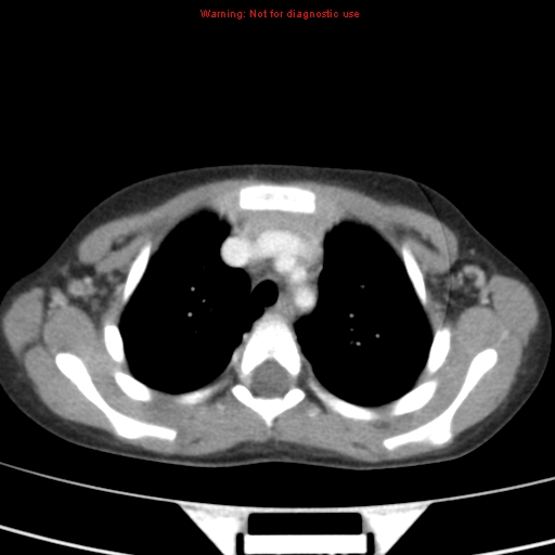
File:Bronchogenic cyst (Radiopaedia 9372-10063 C 3).jpg
Jump to navigation
Jump to search
Bronchogenic_cyst_(Radiopaedia_9372-10063_C_3).jpg (512 × 512 pixels, file size: 72 KB, MIME type: image/jpeg)
Summary:
| Description |
|
| Date | Published: 9th Apr 2010 |
| Source | https://radiopaedia.org/cases/bronchogenic-cyst-2 |
| Author | Hani Makky Al Salam |
| Permission (Permission-reusing-text) |
http://creativecommons.org/licenses/by-nc-sa/3.0/ |
Licensing:
Attribution-NonCommercial-ShareAlike 3.0 Unported (CC BY-NC-SA 3.0)
File history
Click on a date/time to view the file as it appeared at that time.
| Date/Time | Thumbnail | Dimensions | User | Comment | |
|---|---|---|---|---|---|
| current | 17:22, 25 June 2021 |  | 512 × 512 (72 KB) | Fæ (talk | contribs) | Radiopaedia project rID:9372 (batch #5315-42 C3) |
You cannot overwrite this file.
File usage
There are no pages that use this file.
