File:Brown adipose tissue (Radiopaedia 33317).png
Jump to navigation
Jump to search
Size of this preview: 605 × 599 pixels. Other resolutions: 242 × 240 pixels | 484 × 480 pixels | 984 × 975 pixels.
Original file (984 × 975 pixels, file size: 531 KB, MIME type: image/png)
Summary:
- Radiopaedia case ID: 33317
- Image ID: 10234340
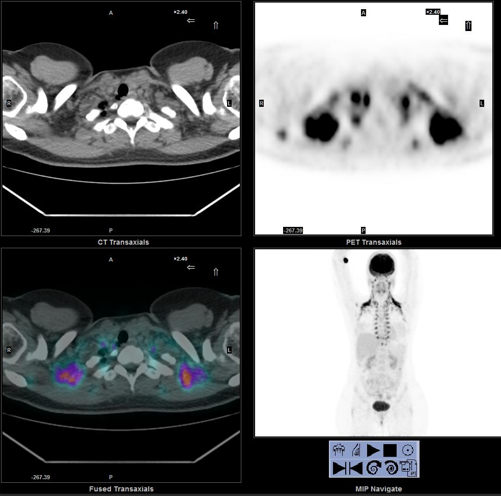
- Study description: 18F-FDG PET/CT
- Modality: Nuclear medicine
- System: Musculoskeletal
- Findings: Symmetric intense FDG uptake in the neck, supraclavicular fossa, paravertebral, axilla and mediastinum, corresponding to fat areas, is shown. These hypermetabolic areas are compatible with activated brown adipose fat (BAT) and shouldn't be confused with malignancy localizations.
- Published: 7th Jan 2015
- Source: https://radiopaedia.org/cases/brown-adipose-tissue
- Author: Anna Margherita Maffione
- Permission: http://creativecommons.org/licenses/by-nc-sa/3.0/
Licensing:
Attribution-NonCommercial-ShareAlike 3.0 Unported (CC BY-NC-SA 3.0)
File history
Click on a date/time to view the file as it appeared at that time.
| Date/Time | Thumbnail | Dimensions | User | Comment | |
|---|---|---|---|---|---|
| current | 09:21, 20 March 2021 | | 984 × 975 (531 KB) | Fæ (talk | contribs) | Radiopaedia project rID:33317 (batch #5176) |
You cannot overwrite this file.
File usage
The following page uses this file:
.png&oldid=8859437)