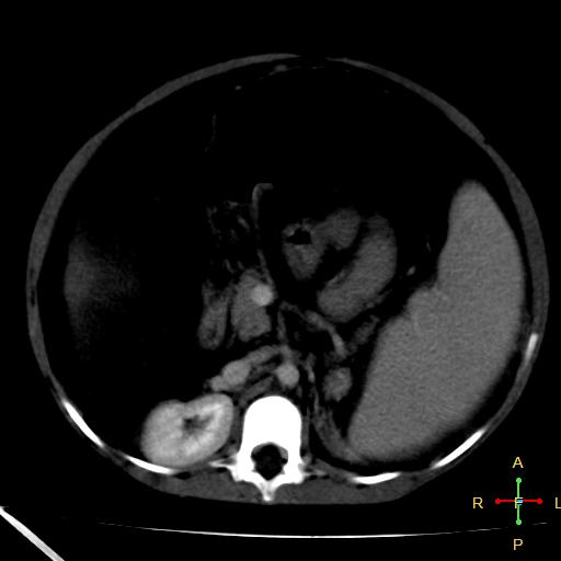
File:Budd-Chiari syndrome (Radiopaedia 23072-23103 B 12).jpg
Jump to navigation
Jump to search
Budd-Chiari_syndrome_(Radiopaedia_23072-23103_B_12).jpg (512 × 512 pixels, file size: 20 KB, MIME type: image/jpeg)
Summary:
| Description |
|
| Date | Published: 20th May 2013 |
| Source | https://radiopaedia.org/cases/budd-chiari-syndrome-1 |
| Author | Ahmed Abdrabou |
| Permission (Permission-reusing-text) |
http://creativecommons.org/licenses/by-nc-sa/3.0/ |
Licensing:
Attribution-NonCommercial-ShareAlike 3.0 Unported (CC BY-NC-SA 3.0)
File history
Click on a date/time to view the file as it appeared at that time.
| Date/Time | Thumbnail | Dimensions | User | Comment | |
|---|---|---|---|---|---|
| current | 19:31, 26 June 2021 |  | 512 × 512 (20 KB) | Fæ (talk | contribs) | Radiopaedia project rID:23072 (batch #5426-28 B12) |
You cannot overwrite this file.
File usage
There are no pages that use this file.
