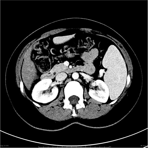
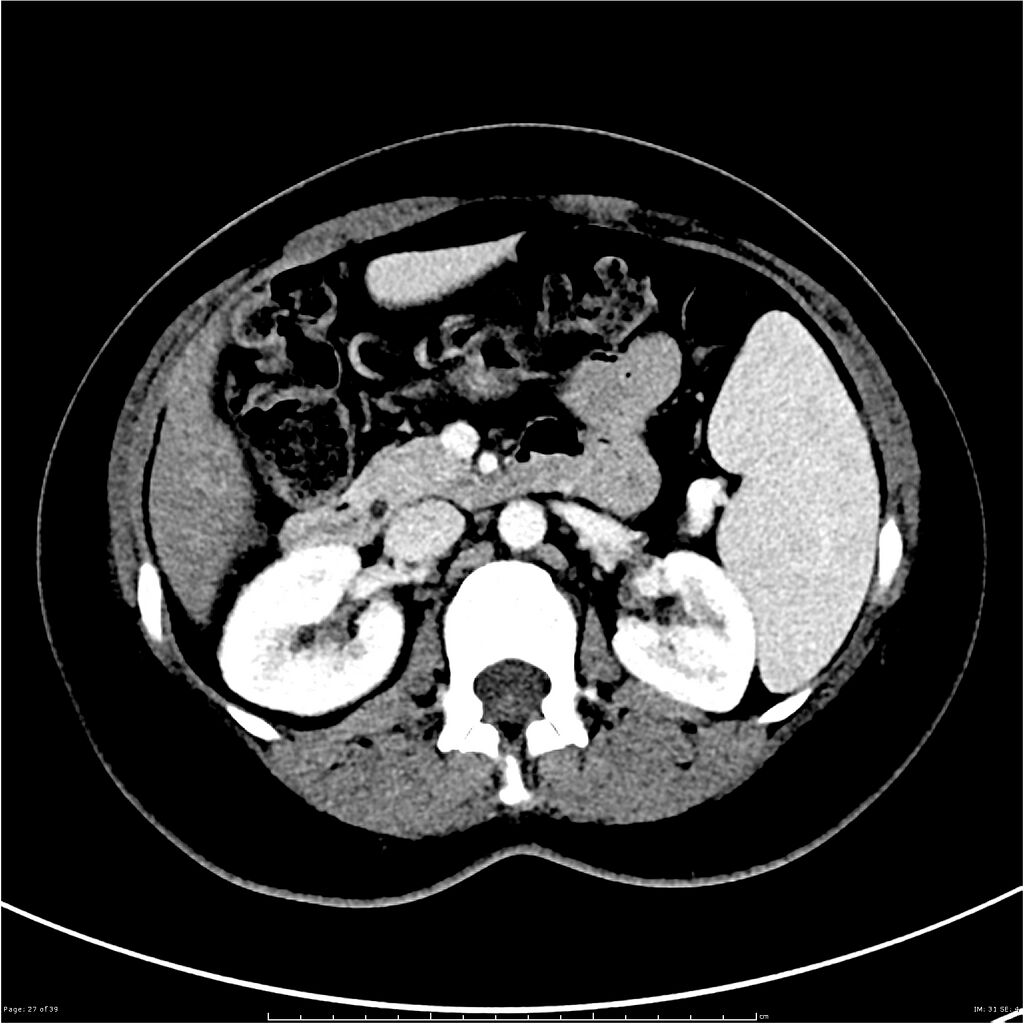
File:Budd-Chiari syndrome (Radiopaedia 27929-28177 Axial liver window 27).JPG
Jump to navigation
Jump to search

Size of this preview: 600 × 600 pixels. Other resolutions: 240 × 240 pixels | 480 × 480 pixels | 768 × 768 pixels | 1,024 × 1,024 pixels | 1,733 × 1,733 pixels.
Original file (1,733 × 1,733 pixels, file size: 636 KB, MIME type: image/jpeg)
Summary:
| Description |
|
| Date | Published: 3rd Mar 2014 |
| Source | https://radiopaedia.org/cases/budd-chiari-syndrome-2 |
| Author | Henry Knipe |
| Permission (Permission-reusing-text) |
http://creativecommons.org/licenses/by-nc-sa/3.0/ |
Licensing:
Attribution-NonCommercial-ShareAlike 3.0 Unported (CC BY-NC-SA 3.0)
File history
Click on a date/time to view the file as it appeared at that time.
| Date/Time | Thumbnail | Dimensions | User | Comment | |
|---|---|---|---|---|---|
| current | 17:40, 26 June 2021 | | 1,733 × 1,733 (636 KB) | Fæ (talk | contribs) | Radiopaedia project rID:27929 (batch #5421-59 B27) |
You cannot overwrite this file.
File usage
There are no pages that use this file.
.JPG&oldid=638562)