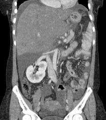
File:Budd-Chiari syndrome (Radiopaedia 56583-63333 D 31).jpg
Jump to navigation
Jump to search

Size of this preview: 533 × 600 pixels. Other resolutions: 213 × 240 pixels | 561 × 631 pixels.
Original file (561 × 631 pixels, file size: 155 KB, MIME type: image/jpeg)
Summary:
| Description |
|
| Date | Published: 24th Nov 2017 |
| Source | https://radiopaedia.org/cases/budd-chiari-syndrome-7 |
| Author | Mostafa El-Feky |
| Permission (Permission-reusing-text) |
http://creativecommons.org/licenses/by-nc-sa/3.0/ |
Licensing:
Attribution-NonCommercial-ShareAlike 3.0 Unported (CC BY-NC-SA 3.0)
File history
Click on a date/time to view the file as it appeared at that time.
| Date/Time | Thumbnail | Dimensions | User | Comment | |
|---|---|---|---|---|---|
| current | 20:08, 26 June 2021 | | 561 × 631 (155 KB) | Fæ (talk | contribs) | Radiopaedia project rID:56583 (batch #5427-214 D31) |
You cannot overwrite this file.
File usage
The following page uses this file:
.jpg&oldid=639545)