File:Burkitt lymphoma HIV (Radiopaedia 32685-33651 A 34).jpg
Jump to navigation
Jump to search
No higher resolution available.
Burkitt_lymphoma_HIV_(Radiopaedia_32685-33651_A_34).jpg (512 × 512 pixels, file size: 36 KB, MIME type: image/jpeg)
Summary:
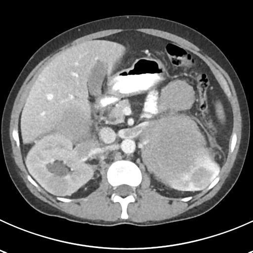
| Description |
|
| Date | Published: 12th Dec 2014 |
| Source | https://radiopaedia.org/cases/burkitt-lymphoma-hiv |
| Author | Frank Gaillard |
| Permission (Permission-reusing-text) |
http://creativecommons.org/licenses/by-nc-sa/3.0/ |
Licensing:
Attribution-NonCommercial-ShareAlike 3.0 Unported (CC BY-NC-SA 3.0)
File history
Click on a date/time to view the file as it appeared at that time.
| Date/Time | Thumbnail | Dimensions | User | Comment | |
|---|---|---|---|---|---|
| current | 03:13, 27 June 2021 | | 512 × 512 (36 KB) | Fæ (talk | contribs) | Radiopaedia project rID:32685 (batch #5463-34 A34) |
You cannot overwrite this file.
File usage
The following page uses this file:
.jpg&oldid=642084)