File:Butterfly glioma (Radiopaedia 76707-88526 Axial SWI 29).jpg

From NC Commons
Jump to navigation Jump to search

Original file(630 × 705 pixels, file size: 54 KB, MIME type: image/jpeg)

Summary:

Description
  • Radiopaedia case ID: 76707
  • Image ID: 52477131
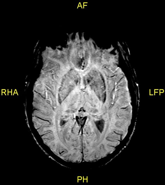
  • Image stack position: 29/60
  • Plane projection: Axial
  • Aux modality: SWI
  • Modality: MRI
  • System: Oncology
  • Findings: Plain MRI Brain {IV contrast (gadolinium) was not given due to our limited experience regarding its safe use in pregnancy}. The scan shows a focal ill-defined heterogeneous mass lesion measuring 3 x 4 x 7 cm in the anterior corpus callosum with extension, on either side of the corpus callosum into the white matter of both frontal lobes (Butterfly lesion). The mass is hypointense on T1 & hyperintense on T2 & FLAIR images. Multiple tiny hemorrhagic foci are seen in the mass on SWI. The mass shows mild diffusion restriction. Moderate vasogenic edema is noted around the lesion in both frontal lobes with extension into the anterior limbs of both internal capsules and into the external capsule-claustrum regions.  Mass effect is noted over the frontal horns of both lateral ventricles; however, no midline shift or hydrocephalus is seen. Remaining brain parenchyma is normal. Based on these MRI features, a few possibilities are high-grade glioma, primary CNS lymphoma, tumefactive demyelination, or an atypical infection like toxoplasmosis.
Date Published: 27th Apr 2020
Source https://radiopaedia.org/cases/butterfly-glioma-2
Author Ammar Ashraf
Permission
(Permission-reusing-text)
http://creativecommons.org/licenses/by-nc-sa/3.0/

Licensing:

Attribution-NonCommercial-ShareAlike 3.0 Unported (CC BY-NC-SA 3.0)

File history

Click on a date/time to view the file as it appeared at that time.

Date/TimeThumbnailDimensionsUserComment
current11:04, 27 June 2021Thumbnail for version as of 11:04, 27 June 2021630 × 705 (54 KB) (talk | contribs)Radiopaedia project rID:76707 (batch #5483-193 G29)

The following page uses this file: