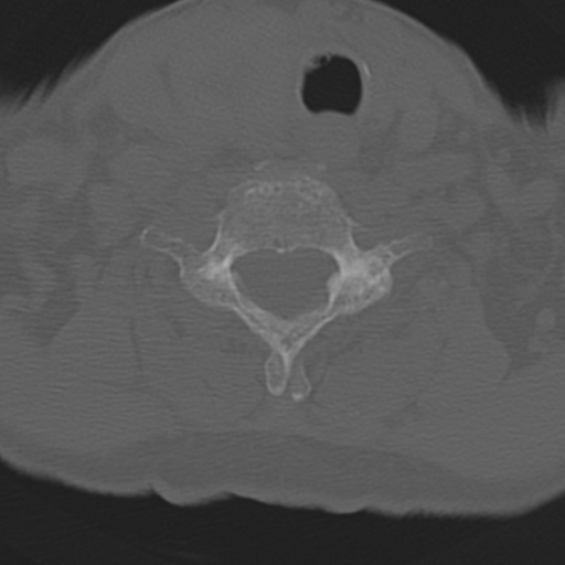
File:C2 fracture with vertebral artery dissection (Radiopaedia 37378-39199 Axial bone window 40).png
Jump to navigation
Jump to search
C2_fracture_with_vertebral_artery_dissection_(Radiopaedia_37378-39199_Axial_bone_window_40).png (512 × 512 pixels, file size: 165 KB, MIME type: image/png)
Summary:
| Description |
|
| Date | Published: 9th Jul 2015 |
| Source | https://radiopaedia.org/cases/c2-fracture-with-vertebral-artery-dissection |
| Author | Craig Hacking |
| Permission (Permission-reusing-text) |
http://creativecommons.org/licenses/by-nc-sa/3.0/ |
Licensing:
Attribution-NonCommercial-ShareAlike 3.0 Unported (CC BY-NC-SA 3.0)
File history
Click on a date/time to view the file as it appeared at that time.
| Date/Time | Thumbnail | Dimensions | User | Comment | |
|---|---|---|---|---|---|
| current | 02:06, 28 June 2021 |  | 512 × 512 (165 KB) | Fæ (talk | contribs) | Radiopaedia project rID:37378 (batch #5508-40 A40) |
You cannot overwrite this file.
File usage
The following page uses this file:
