File:CNS lymphoma (Radiopaedia 36983-38641 Sagittal T1 C+ 20).png

From NC Commons
Jump to navigation Jump to search

CNS_lymphoma_(Radiopaedia_36983-38641_Sagittal_T1_C+_20).png(512 × 512 pixels, file size: 168 KB, MIME type: image/png)

Summary:

Description
  • Radiopaedia case ID: 36983
  • Image ID: 12831623
  • Image stack position: 20/80
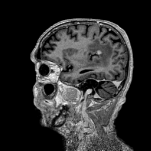
  • Plane projection: Sagittal
  • Aux modality: T1 C+
  • Study description: MRI Brain
  • Study findings: MRI brain demonstrating again the right frontal lobe and left frontoparietal white matter nodular lesions associated with a extensive vasogenic edema. Both lesions show homogeneous and intense contrast enhancement and the left one a mild hemorrhagic posterior component. The study also evidences a similar enhanced nodular lesion in the left cerebral peduncle.
  • Modality: MRI
  • System: Central Nervous System
  • Findings: CT brain shows two hyperdense nodular deep white matter lesions located in the right corona radiata region and in the left frontoparietal transition, both associated with an extensive hypoattenuating area surrounding them and that may correspond to a vasogenic edema. The mass effect is more pronounced in the right where it causes a mild right lateral ventricle compression; bilateral mild sulcal effacement. No midline shift. It is also notated a small not well-defined hypoattenuating area in the left cerebral peduncle.
Date Published: 21st May 2015
Source https://radiopaedia.org/cases/cns-lymphoma-8
Author RMH Neuropathology
Permission
(Permission-reusing-text)
http://creativecommons.org/licenses/by-nc-sa/3.0/

Licensing:

Attribution-NonCommercial-ShareAlike 3.0 Unported (CC BY-NC-SA 3.0)

File history

Click on a date/time to view the file as it appeared at that time.

Date/TimeThumbnailDimensionsUserComment
current08:45, 28 August 2021Thumbnail for version as of 08:45, 28 August 2021512 × 512 (168 KB) (talk | contribs)Radiopaedia project rID:36983 (batch #8450-140 F20)

The following page uses this file:

Metadata