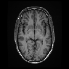
File:CNS tuberculomas (Radiopaedia 72245-82766 Axial T1 35).jpg
Jump to navigation
Jump to search
CNS_tuberculomas_(Radiopaedia_72245-82766_Axial_T1_35).jpg (230 × 230 pixels, file size: 20 KB, MIME type: image/jpeg)
Summary:
| Description |
|
| Date | Published: 13th Dec 2019 |
| Source | https://radiopaedia.org/cases/cns-tuberculomas-1 |
| Author | Dalia Ibrahim |
| Permission (Permission-reusing-text) |
http://creativecommons.org/licenses/by-nc-sa/3.0/ |
Licensing:
Attribution-NonCommercial-ShareAlike 3.0 Unported (CC BY-NC-SA 3.0)
File history
Click on a date/time to view the file as it appeared at that time.
| Date/Time | Thumbnail | Dimensions | User | Comment | |
|---|---|---|---|---|---|
| current | 18:04, 28 August 2021 |  | 230 × 230 (20 KB) | Fæ (talk | contribs) | Radiopaedia project rID:72245 (batch #8460-75 C35) |
You cannot overwrite this file.
File usage
The following page uses this file:
