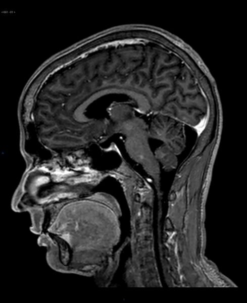
File:CNS tuberculosis (Radiopaedia 40083-42588 Sagittal T1 C+ 63).jpg
Jump to navigation
Jump to search

Size of this preview: 489 × 600 pixels. Other resolutions: 196 × 240 pixels | 391 × 480 pixels | 626 × 768 pixels | 835 × 1,024 pixels | 1,504 × 1,844 pixels.
Original file (1,504 × 1,844 pixels, file size: 232 KB, MIME type: image/jpeg)
Summary:
| Description |
|
| Date | Published: 6th Oct 2015 |
| Source | https://radiopaedia.org/cases/cns-tuberculosis |
| Author | Mark Hall |
| Permission (Permission-reusing-text) |
http://creativecommons.org/licenses/by-nc-sa/3.0/ |
Licensing:
Attribution-NonCommercial-ShareAlike 3.0 Unported (CC BY-NC-SA 3.0)
File history
Click on a date/time to view the file as it appeared at that time.
| Date/Time | Thumbnail | Dimensions | User | Comment | |
|---|---|---|---|---|---|
| current | 19:43, 28 August 2021 |  | 1,504 × 1,844 (232 KB) | Fæ (talk | contribs) | Radiopaedia project rID:40083 (batch #8461-179 D63) |
You cannot overwrite this file.
File usage
The following page uses this file: