File:CNS vasculitis (Radiopaedia 55715-62264 Sagittal T1 C+ 98).png

From NC Commons
Jump to navigation Jump to search

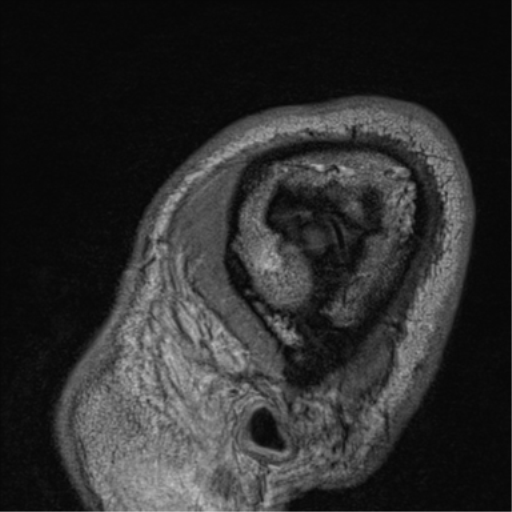
CNS_vasculitis_(Radiopaedia_55715-62264_Sagittal_T1_C+_98).png(512 × 512 pixels, file size: 129 KB, MIME type: image/png)

Summary:

Description
  • Radiopaedia case ID: 55715
  • Image ID: 32824956
  • Image stack position: 98/99
  • Plane projection: Sagittal
  • Aux modality: T1 C+
  • Study findings: There is widespread long segment concentric vessel wall thickening and hyperenhancement throughout the brain, involving particularly the M2, M3 and M4 MCA branches bilaterally, bilateral PCAs and right medial lenticulostriate vessels. Known focal stenosis of the superior M2 division of the left MCA. Occluded M3 branch of left inferior M2 division with focal T1 hyperintensity in the left sylvian fissure. Occluded M3 branch of the right inferior M2 division. Known subacute infarct in the right basal ganglia. Conclusion: Extensive bilateral hemisphere long segmental vessel wall thickening and hyperenhancement in keeping with vasculitis. Secondary and primary vasculitis both need to be considered.
  • Modality: MRI
  • System: Central Nervous System
  • Findings: Hypoattenuation involving the right caudate and lentiform nucleus is favored to represent a combination of chronic and subacute ischemia.
Date Published: 22nd Sep 2017
Source https://radiopaedia.org/cases/cns-vasculitis-5
Author Ernest Lekgabe
Permission
(Permission-reusing-text)
http://creativecommons.org/licenses/by-nc-sa/3.0/

Licensing:

Attribution-NonCommercial-ShareAlike 3.0 Unported (CC BY-NC-SA 3.0)

File history

Click on a date/time to view the file as it appeared at that time.

Date/TimeThumbnailDimensionsUserComment
current21:49, 28 August 2021Thumbnail for version as of 21:49, 28 August 2021512 × 512 (129 KB) (talk | contribs)Radiopaedia project rID:55715 (batch #8462-273 C98)

The following page uses this file:

Metadata