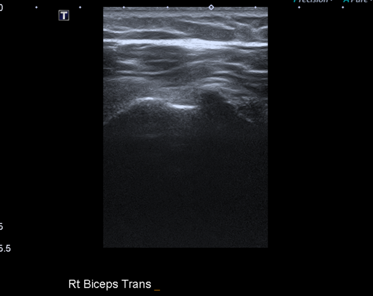
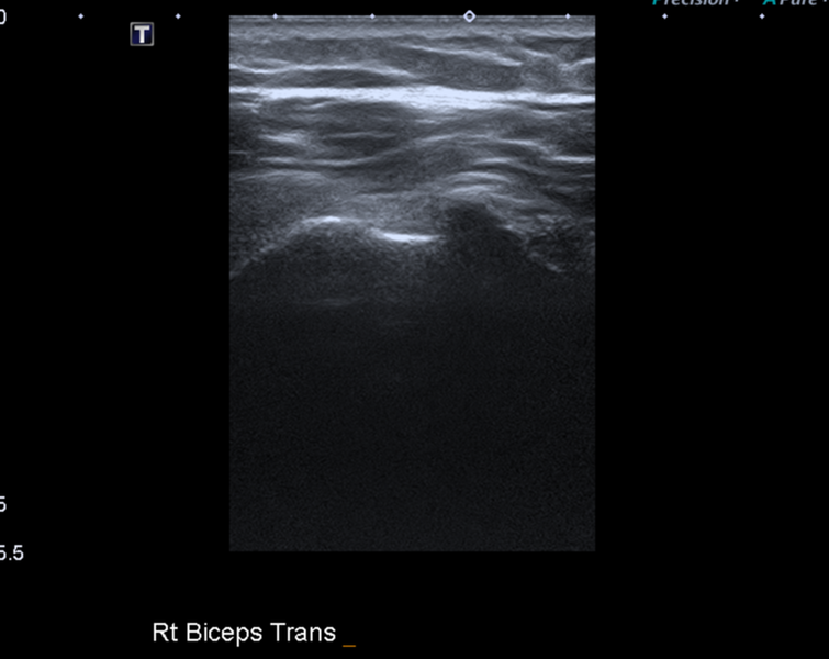
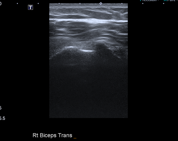
File:Calcific tendinitis (ruptured) (Radiopaedia 56393-63053 A 1).png
Jump to navigation
Jump to search
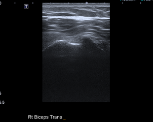
Size of this preview: 755 × 600 pixels. Other resolutions: 302 × 240 pixels | 604 × 480 pixels | 768 × 610 pixels.
Original file (768 × 610 pixels, file size: 201 KB, MIME type: image/png)
Summary:
| Description |
|
| Date | Published: 12th Nov 2017 |
| Source | https://radiopaedia.org/cases/calcific-tendinitis-ruptured |
| Author | Bruno Di Muzio |
| Permission (Permission-reusing-text) |
http://creativecommons.org/licenses/by-nc-sa/3.0/ |
Licensing:
Attribution-NonCommercial-ShareAlike 3.0 Unported (CC BY-NC-SA 3.0)
File history
Click on a date/time to view the file as it appeared at that time.
| Date/Time | Thumbnail | Dimensions | User | Comment | |
|---|---|---|---|---|---|
| current | 00:08, 30 June 2021 | | 768 × 610 (201 KB) | Fæ (talk | contribs) | Radiopaedia project rID:56393 (batch #5725-1 A1) |
You cannot overwrite this file.
File usage
There are no pages that use this file.
_(Radiopaedia_56393-63053_A_1).png&oldid=663515)