File:Calcific tendinitis of the longus colli muscle (Radiopaedia 36426).jpg
Jump to navigation
Jump to search

Size of this preview: 527 × 599 pixels. Other resolutions: 211 × 240 pixels | 422 × 480 pixels | 675 × 768 pixels | 1,109 × 1,261 pixels.
Original file (1,109 × 1,261 pixels, file size: 257 KB, MIME type: image/jpeg)
Summary:
- Radiopaedia case ID: 36426
- Image ID: 30651
- Study findings: Calcific tendinitis of the longus colli muscle
- Modality: Annotated image
- System: Musculoskeletal
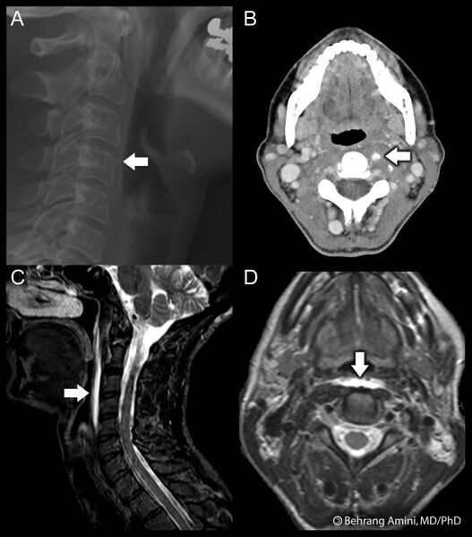
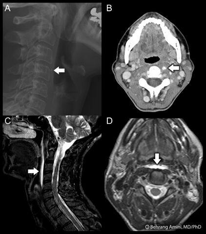
- Findings: Calcific tendinitis of the longus colli muscle
(A) a small calcification anterior to the C4 vertebral body on the radiograph (arrow). (B) the amorphous calcification is better evaluated on the CT (arrow). sagittal STIR (C) and coronal T2-weighted (D) images show edema anterior to the longus colli muscle.
- Published: 30th Apr 2015
- Source: https://radiopaedia.org/cases/calcific-tendinitis-of-the-longus-colli-muscle-1
- Author: Behrang Amini
- Permission: http://creativecommons.org/licenses/by-nc-sa/3.0/
Licensing:
Attribution-NonCommercial-ShareAlike 3.0 Unported (CC BY-NC-SA 3.0)
| This file is ineligible for copyright and therefore in the public domain, because it is a technical image created as part of a standard medical diagnostic procedure. No creative element rising above the threshold of originality was involved in its production.
|
 |
File history
Click on a date/time to view the file as it appeared at that time.
| Date/Time | Thumbnail | Dimensions | User | Comment | |
|---|---|---|---|---|---|
| current | 10:23, 20 March 2021 | | 1,109 × 1,261 (257 KB) | Fæ (talk | contribs) | Radiopaedia project rID:36426 (batch #5534) |
You cannot overwrite this file.
File usage
There are no pages that use this file.
.jpg&oldid=9756163)