File:Calcified abdominal aortic aneurysm on pelvic radiograph (Radiopaedia 63020).jpg
Jump to navigation
Jump to search
Size of this preview: 621 × 599 pixels. Other resolutions: 249 × 240 pixels | 497 × 480 pixels | 796 × 768 pixels | 1,061 × 1,024 pixels | 2,122 × 2,048 pixels | 2,971 × 2,868 pixels.
Original file (2,971 × 2,868 pixels, file size: 1.05 MB, MIME type: image/jpeg)
Summary:
- Radiopaedia case ID: 63020
- Image ID: 42377935
- Modality: X-ray
- System: Vascular
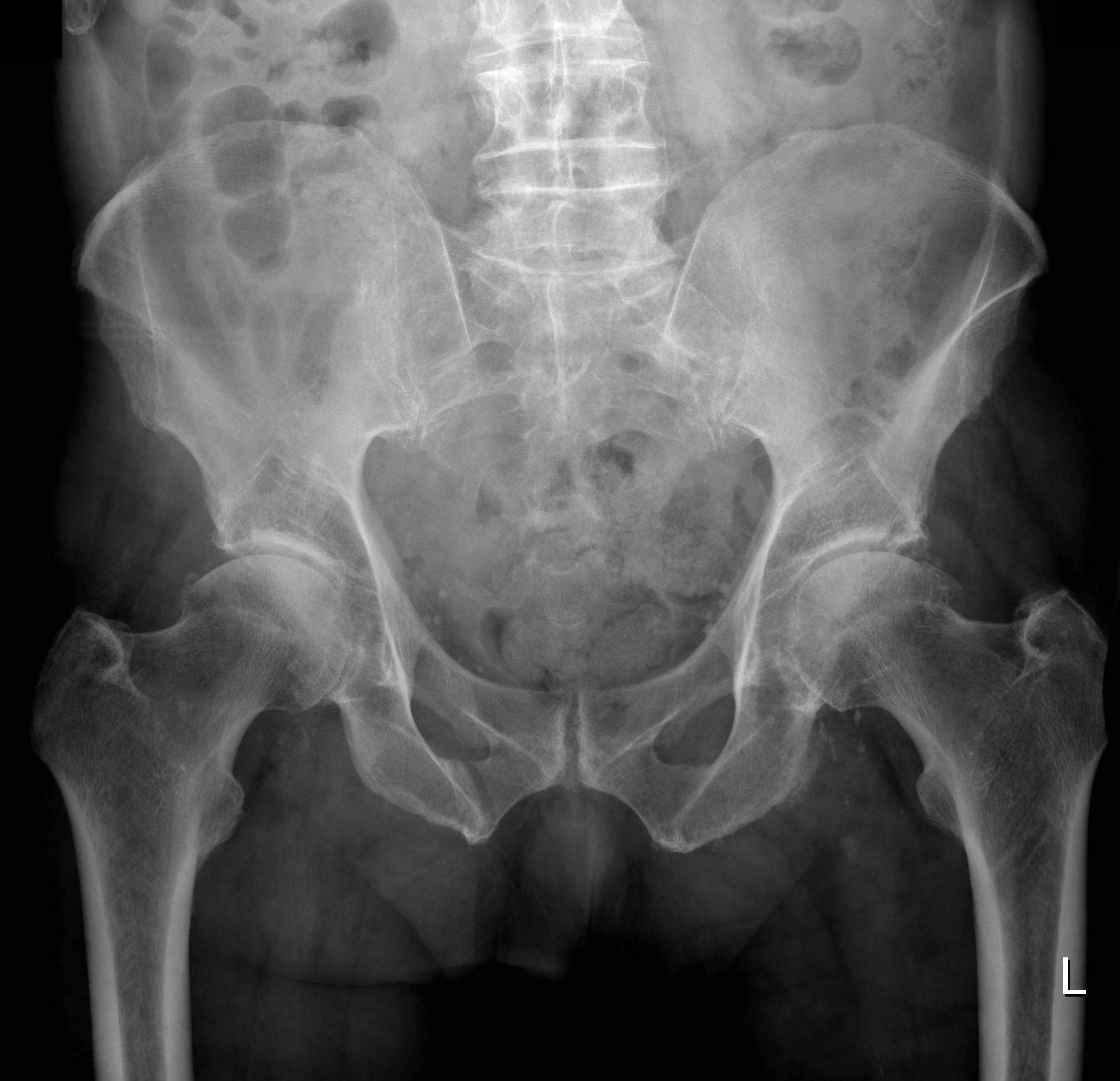
- Findings: Degenerative changes of both hips. No fracture or mal-alignment. SI joints are normal. Projected over the lower lumbar vertebrae, the abdominal aorta is calcified and aneurysmal.
- Published: 10th Sep 2018
- Source: https://radiopaedia.org/cases/calcified-abdominal-aortic-aneurysm-on-pelvic-radiograph
- Author: Craig Hacking
- Permission: http://creativecommons.org/licenses/by-nc-sa/3.0/
Licensing:
Attribution-NonCommercial-ShareAlike 3.0 Unported (CC BY-NC-SA 3.0)
File history
Click on a date/time to view the file as it appeared at that time.
| Date/Time | Thumbnail | Dimensions | User | Comment | |
|---|---|---|---|---|---|
| current | 10:29, 20 March 2021 | | 2,971 × 2,868 (1.05 MB) | Fæ (talk | contribs) | Radiopaedia project rID:63020 (batch #5563) |
You cannot overwrite this file.
File usage
The following page uses this file:
.jpg&oldid=8859374)